The CYTH1 Antibody (CAB15351) is a high-quality antibody developed for reliable detection and analysis of target proteins. This antibody, produced in rabbits, exhibits high reactivity with human samples and has been validated for use in Western blot applications. By binding specifically to the CYTH1 protein, this antibody enables the detection and analysis of CYTH1 in various cell types.CYTH1, also known as Cytohesin-1, plays a crucial role in regulating vesicular trafficking and intracellular signaling pathways. Dysregulation of CYTH1 has been implicated in various diseases, including cancer, neurodegenerative disorders, and infectious diseases.
This antibody is validated for use in WB, IF/ICC, ELISA applications and has demonstrated reactivity against Human, Mouse, Rat samples.
Product Name:
CYTH1 Antibody
SKU:
CAB15351
Size:
20μL, 100μL
Reactivity:
Human, Mouse, Rat
Conjugate:
Unconjugated
Immunogen:
Recombinant protein (or fragment).This information is considered to be commercially sensitive.
Sequence:
MEED DSYV PSDL TAEE RQEL ENIR RRKQ ELLA DIQR LKDE IAEV ANEI ENLG STEE RKNM QRNK QVAM GR
Tested Applications:
WBIF/ICCELISA
Recommended Dilution:
WB
1:200 - 1:2000
IF/ICC
1:50 - 1:200
ELISA
Recommended starting concentration is 1 μg/mL. Please optimize the concentration based on your specific assay requirements.
Synonyms:
B2-1, SEC7, PSCD1, D17S811E, CYTOHESIN-1, CYTH1
Positive Sample:
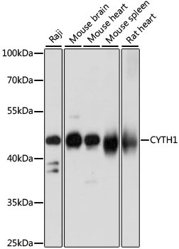
Raji, Mouse brain, Mouse heart, Mouse spleen, Rat heart
The protein encoded by this gene is a member of the PSCD family. Members of this family have identical structural organization that consists of an N-terminal coiled-coil motif, a central Sec7 domain, and a C-terminal pleckstrin homology (PH) domain. The coiled-coil motif is involved in homodimerization, the Sec7 domain contains guanine-nucleotide exchange protein activity, and the PH domain interacts with phospholipids and is responsible for association of PSCDs with membranes. Members of this family appear to mediate the regulation of protein sorting and membrane trafficking. This gene is highly expressed in natural killer and peripheral T cells, and regulates the adhesiveness of integrins at the plasma membrane of lymphocytes. A pseudogene of this gene has been defined on the X chromosome. Alternative splicing results in multiple transcript variants.
Purification Method
Affinity purification
Gene ID
9267
RRID
AB_2762253
Buffer Information
Store at -20℃. Avoid freeze / thaw cycles. Buffer: PBS with 0.01% thimerosal,50% glycerol,pH7.3.
Western blot analysis of various lysates using CYTH1 Rabbit pAb (CAB15351) at 1:1000 dilution. Secondary antibody: HRP-conjugated Goat anti-Rabbit IgG (H+L) (CABS014) at 1:10000 dilution. Lysates/proteins: 25μg per lane. Blocking buffer: 3% nonfat dry milk in TBST. Detection: ECL Basic Kit (AbGn00020). Exposure time: 5s.
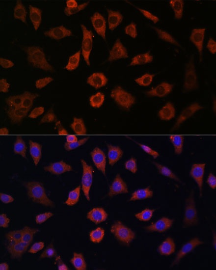
Immunofluorescence analysis of L929 cells using CYTH1 Rabbit pAb (CAB15351) at dilution of 1:100. Secondary antibody: Cy3-conjugated Goat anti-Rabbit IgG (H+L) (CABS007) at 1:500 dilution. Blue: DAPI for nuclear staining.