The CDA Antibody (CAB2006) is a high-quality antibody developed for reliable detection and analysis of target proteins. This antibody, produced in rabbits, specifically targets cytidine deaminase and is highly reactive with human samples. It has been validated for use in Western blot applications, allowing for the detection and analysis of cytidine deaminase in various cell types.Cytidine deaminase plays a critical role in the conversion of cytidine to uridine, a process that is crucial for DNA and RNA synthesis. Dysregulation of cytidine deaminase has been linked to various diseases, including cancer and autoimmune disorders.
This antibody is validated for use in WB, IF/ICC, ELISA applications and has demonstrated reactivity against Human, Mouse, Rat samples.
Product Name:
CDA Antibody
SKU:
CAB2006
Size:
20μL, 100μL
Reactivity:
Human, Mouse, Rat
Conjugate:
Unconjugated
Immunogen:
Recombinant protein (or fragment).This information is considered to be commercially sensitive.
Recommended starting concentration is 1 μg/mL. Please optimize the concentration based on your specific assay requirements.
Synonyms:
CDD, CDA
Positive Sample:
Mouse kidney
Cellular Localization:
Cytosol, Extracellular Region.
Calculated MW:
16kDa
Observed MW:
16kDa
This gene encodes an enzyme involved in pyrimidine salvaging. The encoded protein forms a homotetramer that catalyzes the irreversible hydrolytic deamination of cytidine and deoxycytidine to uridine and deoxyuridine, respectively. It is one of several deaminases responsible for maintaining the cellular pyrimidine pool. Mutations in this gene are associated with decreased sensitivity to the cytosine nucleoside analogue cytosine arabinoside used in the treatment of certain childhood leukemias.
Purification Method
Affinity purification
Gene ID
978
RRID
AB_2764030
Buffer Information
Store at -20℃. Avoid freeze / thaw cycles. Buffer: PBS containing 50% glycerol, preserved with proclin300 or sodium azide, pH 7.3.
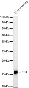
Western blot analysis of lysates from Mouse kidney, using CDA Rabbit pAb (CAB2006) at 1:400 dilution. Secondary antibody: HRP-conjugated Goat anti-Rabbit IgG (H+L) (CABS014) at 1:10000 dilution. Lysates/proteins: 25μg per lane. Blocking buffer: 3% nonfat dry milk in TBST. Detection: ECL Basic Kit (AbGn00020). Exposure time: 180s.
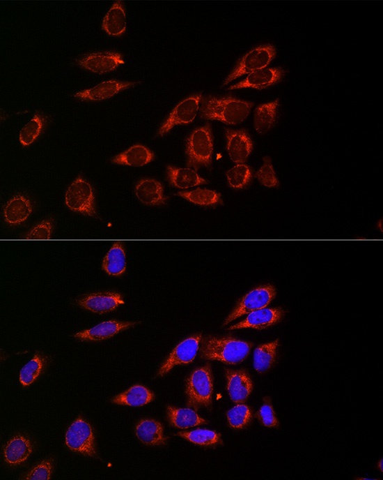
Immunofluorescence analysis of HeLa cells using CDA Rabbit pAb (CAB2006) at dilution of 1:20 (40x lens). Secondary antibody: Cy3-conjugated Goat anti-Rabbit IgG (H+L) (CABS007) at 1:500 dilution. Blue: DAPI for nuclear staining.
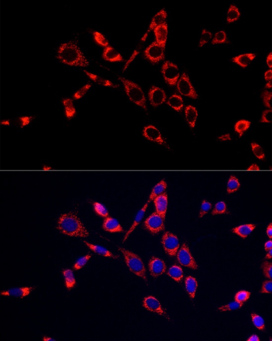
Immunofluorescence analysis of NIH/3T3 cells using CDA Rabbit pAb (CAB2006) at dilution of 1:20 (40x lens). Secondary antibody: Cy3-conjugated Goat anti-Rabbit IgG (H+L) (CABS007) at 1:500 dilution. Blue: DAPI for nuclear staining.
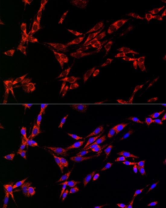
Immunofluorescence analysis of PC-12 cells using CDA Rabbit pAb (CAB2006) at dilution of 1:20 (40x lens). Secondary antibody: Cy3-conjugated Goat anti-Rabbit IgG (H+L) (CABS007) at 1:500 dilution. Blue: DAPI for nuclear staining.