The DCT Antibody (CAB16041) is a high-quality antibody developed for reliable detection and analysis of target proteins. This antibody, raised in rabbits, is highly specific and reacts well with human samples, making it suitable for various applications such as Western blot and immunohistochemistry.DCT, also known as dopachrome tautomerase, plays a crucial role in the melanogenesis pathway by catalyzing the conversion of dopachrome to dihydroxyindole carboxylic acid (DHICA). This process is essential for the production of eumelanin, the dark pigment responsible for determining skin, hair, and eye color.
This antibody is validated for use in WB, ELISA applications and has demonstrated reactivity against Human, Mouse samples.
Product Name:
DCT Antibody
SKU:
CAB16041
Size:
20μL, 100μL
Reactivity:
Human, Mouse
Conjugate:
Unconjugated
Immunogen:
Recombinant protein (or fragment).This information is considered to be commercially sensitive.
Recommended starting concentration is 1 μg/mL. Please optimize the concentration based on your specific assay requirements.
Synonyms:
OCA8, TRP-2, TYRP2, DCT
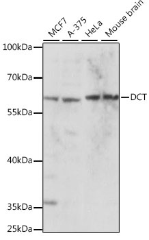
Positive Sample:
MCF7, A-375, HeLa, Mouse brain
Cellular Localization:
Melanosome Membrane, Single-Pass Type I Membrane Protein.
Calculated MW:
59kDa
Observed MW:
59kDa
Predicted to enable dopachrome isomerase activity. Involved in response to blue light. Located in intracellular membrane-bounded organelle and plasma membrane. Implicated in oculocutaneous albinism.
Purification Method
Affinity purification
Gene ID
1638
RRID
AB_2763480
Buffer Information
Store at -20℃. Avoid freeze / thaw cycles. Buffer: PBS with 0.01% thimerosal,50% glycerol,pH7.3.
Western blot analysis of various lysates using DCT Rabbit pAb (CAB16041) at 1:1000 dilution. Secondary antibody: HRP-conjugated Goat anti-Rabbit IgG (H+L) (CABS014) at 1:10000 dilution. Lysates/proteins: 25μg per lane. Blocking buffer: 3% nonfat dry milk in TBST. Detection: ECL Basic Kit (AbGn00020). Exposure time: 10s.