The DDX1 Antibody (CAB6575) is a high-quality antibody developed for reliable detection and analysis of target proteins. This antibody, generated in rabbits, is highly specific to DDX1 and is suitable for use in various applications such as Western blot and immunohistochemistry.DDX1 is a member of the DEAD-box RNA helicase family and is known to play a role in RNA processing and RNA virus replication. By targeting DDX1 with this antibody, researchers can gain valuable insights into the function and regulation of this important protein in cellular processes such as RNA splicing, translation, and RNA virus replication.
This antibody is validated for use in WB, IHC-P, IF/ICC, IP, ELISA applications and has demonstrated reactivity against Human, Mouse, Rat samples.
Product Name:
DDX1 Antibody
SKU:
CAB6575
Size:
20μL, 100μL
Reactivity:
Human, Mouse, Rat
Conjugate:
Unconjugated
Immunogen:
Recombinant protein (or fragment).This information is considered to be commercially sensitive.
0.5μg-4μg antibody for 200μg-400μg extracts of whole cells
ELISA
Recommended starting concentration is 1 μg/mL. Please optimize the concentration based on your specific assay requirements.
Synonyms:
DBP-RB, UKVH5d, DDX1
Positive Sample:
293F
Cellular Localization:
Cytoplasm, Cytoplasmic Granule, Nucleus.
Calculated MW:
82kDa
Observed MW:
83kDa
DEAD box proteins, characterized by the conserved motif Asp-Glu-Ala-Asp (DEAD), are putative RNA helicases. They are implicated in a number of cellular processes involving alteration of RNA secondary structure such as translation initiation, nuclear and mitochondrial splicing, and ribosome and spliceosome assembly. Based on their distribution patterns, some members of this family are believed to be involved in embryogenesis, spermatogenesis, and cellular growth and division. This gene encodes a DEAD box protein that acts as an ATP-dependent RNA helicase that has been found to promote coronaviruses replication.
Purification Method
Affinity purification
Gene ID
1653
RRID
AB_2767169
Buffer Information
Store at -20℃. Avoid freeze / thaw cycles. Buffer: PBS containing 50% glycerol, preserved with proclin300 or sodium azide, pH 7.3.
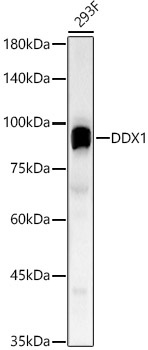
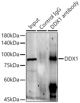
Western blot analysis of lysates from 293F cells, using DDX1 Rabbit pAb (CAB6575) at 1:2000 dilution. Secondary antibody: HRP-conjugated Goat anti-Rabbit IgG (H+L) (CABS014) at 1:10000 dilution. Lysates/proteins: 25μg per lane. Blocking buffer: 3% nonfat dry milk in TBST. Detection: ECL Basic Kit (AbGn00020). Exposure time: 60s.
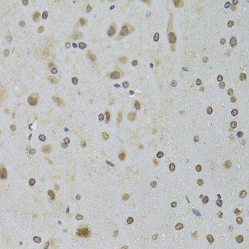
Immunohistochemistry analysis of paraffin-embedded Rat brain using DDX1 Rabbit pAb (CAB6575) at dilution of 1:100 (40x lens). Microwave antigen retrieval performed with 0.01M PBS Buffer (pH 7.2) prior to IHC staining.
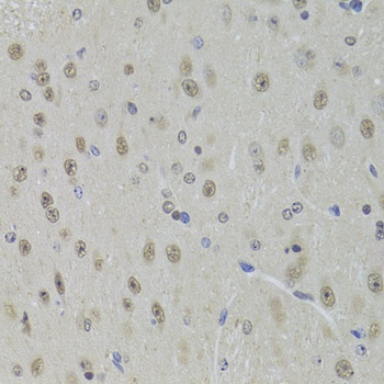
Immunohistochemistry analysis of paraffin-embedded Mouse brain using DDX1 Rabbit pAb (CAB6575) at dilution of 1:100 (40x lens). Microwave antigen retrieval performed with 0.01M PBS Buffer (pH 7.2) prior to IHC staining.
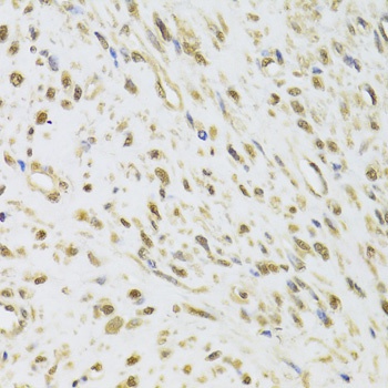
Immunohistochemistry analysis of paraffin-embedded Human leiomyoma of uterus using DDX1 Rabbit pAb (CAB6575) at dilution of 1:100 (40x lens). Microwave antigen retrieval performed with 0.01M PBS Buffer (pH 7.2) prior to IHC staining.
Immunoprecipitation analysis of 300 μg extracts of 293F cells using 3 μg DDX1 Rabbit pAb (CAB6575). Western blot was performed from the immunoprecipitate using DDX1 Rabbit pAb (CAB6575) at a dilition of 1:2000.