The DHFR Antibody (CAB1607) is a high-quality antibody developed for reliable detection and analysis of target proteins. This antibody, produced in rabbits, is highly specific to human samples and has been validated for use in Western blot applications. By binding to the DHFR protein, this antibody allows for accurate detection and analysis in a variety of cell types, making it ideal for studies in cancer research, pharmacology, and drug development.DHFR is a critical enzyme in the folate pathway, playing a key role in the synthesis of nucleotides needed for DNA replication and cell growth. Dysregulation of DHFR has been linked to cancer development and resistance to certain chemotherapy drugs, highlighting the importance of studying this enzyme in disease progression.
This antibody is validated for use in WB, IF/ICC, ELISA applications and has demonstrated reactivity against Human, Mouse samples.
Product Name:
DHFR Antibody
SKU:
CAB1607
Size:
20μL, 100μL
Reactivity:
Human, Mouse
Conjugate:
Unconjugated
Immunogen:
Recombinant protein (or fragment).This information is considered to be commercially sensitive.
Dihydrofolate reductase converts dihydrofolate into tetrahydrofolate, a methyl group shuttle required for the de novo synthesis of purines, thymidylic acid, and certain amino acids. While the functional dihydrofolate reductase gene has been mapped to chromosome 5, multiple intronless processed pseudogenes or dihydrofolate reductase-like genes have been identified on separate chromosomes. Dihydrofolate reductase deficiency has been linked to megaloblastic anemia. Several transcript variants encoding different isoforms have been found for this gene.
Purification Method
Affinity purification
Gene ID
1719
RRID
AB_2763510
Buffer Information
Store at -20℃. Avoid freeze / thaw cycles. Buffer: PBS containing 50% glycerol, preserved with proclin300 or sodium azide, pH 7.3.
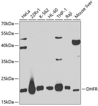
Western blot analysis of various lysates using DHFR Rabbit pAb (CAB1607) at 1:1000 dilution. Secondary antibody: HRP-conjugated Goat anti-Rabbit IgG (H+L) (CABS014) at 1:10000 dilution. Lysates/proteins: 25μg per lane. Blocking buffer: 3% nonfat dry milk in TBST.
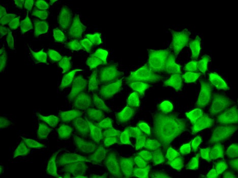
Immunofluorescence analysis of A-549 cells using DHFR Rabbit pAb (CAB1607).Secondary antibody: Cy3-conjugated Goat anti-Rabbit IgG (H+L) (CABS007) at 1:500 dilution.