The DRG2 Antibody (CAB17476) is a high-quality antibody developed for reliable detection and analysis of target proteins. This antibody, produced in rabbits, exhibits high reactivity with human samples and has been rigorously validated for use in Western blotting applications.The DRG2 Polyclonal Antibody specifically binds to the DRG2 protein, enabling precise detection and analysis in a variety of cell types. Its versatility makes it an ideal tool for researchers in the fields of cell biology and cancer research, allowing for detailed exploration of DRG2 function and its potential implications in disease processes.
This antibody is validated for use in WB, IF/ICC, ELISA applications and has demonstrated reactivity against Human, Mouse, Rat samples.
Product Name:
DRG2 Antibody
SKU:
CAB17476
Size:
20μL, 100μL
Reactivity:
Human, Mouse, Rat
Conjugate:
Unconjugated
Immunogen:
Recombinant protein (or fragment).This information is considered to be commercially sensitive.
Recommended starting concentration is 1 μg/mL. Please optimize the concentration based on your specific assay requirements.
Synonyms:
DRG2
Positive Sample:
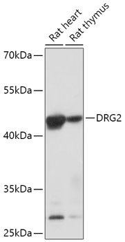
Rat heart, Rat thymus
Cellular Localization:
Cytoplasm, Cytosol, Nucleoplasm, Nucleus.
Calculated MW:
41kDa
Observed MW:
45kDa
This gene encodes a GTP-binding protein known to function in the regulation of cell growth and differentiation. Read-through transcripts containing this gene and a downstream gene have been identified, but they are not thought to encode a fusion protein. This gene is located within the Smith-Magenis syndrome region on chromosome 17.
Purification Method
Affinity purification
Gene ID
1819
RRID
AB_2769227
Buffer Information
Store at -20℃. Avoid freeze / thaw cycles. Buffer: PBS with 0.01% thimerosal,50% glycerol,pH7.3.
Western blot analysis of various lysates using DRG2 Rabbit pAb (CAB17476) at 1:1000 dilution. Secondary antibody: HRP-conjugated Goat anti-Rabbit IgG (H+L) (CABS014) at 1:10000 dilution. Lysates/proteins: 25μg per lane. Blocking buffer: 3% nonfat dry milk in TBST. Detection: ECL Basic Kit (AbGn00020). Exposure time: 60s.
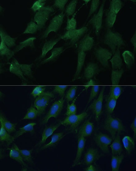
Immunofluorescence analysis of C6 cells using DRG2 Rabbit pAb (CAB17476) at dilution of 1:100. Secondary antibody: Cy3-conjugated Goat anti-Rabbit IgG (H+L) (CABS007) at 1:500 dilution. Blue: DAPI for nuclear staining.