The DUSP1/MKP1 Antibody (CAB2919) is a high-quality antibody developed for reliable detection and analysis of target proteins. DUSP1, also known as MAP kinase phosphatase-1 (MKP-1), plays a crucial role in the regulation of cellular signaling pathways involved in inflammation, immune response, and cell proliferation. This antibody, raised in rabbits, exhibits high reactivity with human samples and has been validated for use in Western blot applications. By specifically binding to DUSP1, researchers can detect and analyze the protein in various cell types, making it ideal for studies in immunology, cancer research, and the development of therapeutics targeting DUSP1.
This antibody is validated for use in WB, ELISA applications and has demonstrated reactivity against Human, Mouse, Rat samples.
Product Name:
DUSP1/MKP1 Antibody
SKU:
CAB2919
Size:
20μL, 100μL
Reactivity:
Human, Mouse, Rat
Conjugate:
Unconjugated
Immunogen:
Recombinant protein (or fragment).This information is considered to be commercially sensitive.
Recommended starting concentration is 1 μg/mL. Please optimize the concentration based on your specific assay requirements.
Synonyms:
HVH1, MKP1, CL100, MKP-1, PTPN10, DUSP1/MKP1
Positive Sample:
Rat brain
Cellular Localization:
Nucleus.
Calculated MW:
39kDa
Observed MW:
40kDa
The protein encoded by this gene is a phosphatase with dual specificity for tyrosine and threonine. The encoded protein can dephosphorylate MAP kinase MAPK1/ERK2, which results in its involvement in several cellular processes. This protein appears to play an important role in the human cellular response to environmental stress as well as in the negative regulation of cellular proliferation. Finally, the encoded protein can make some solid tumors resistant to both chemotherapy and radiotherapy, making it a target for cancer therapy.
Purification Method
Affinity purification
Gene ID
1843
RRID
AB_2764738
Buffer Information
Store at -20℃. Avoid freeze / thaw cycles. Buffer: PBS containing 50% glycerol, preserved with proclin300 or sodium azide, pH 7.3.
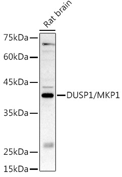
Western blot analysis of lysates from Rat brain, using DUSP1/MKP1 Rabbit pAb (CAB2919) at 1:500 dilution. Secondary antibody: HRP-conjugated Goat anti-Rabbit IgG (H+L) (CABS014) at 1:10000 dilution. Lysates/proteins: 25μg per lane. Blocking buffer: 3% nonfat dry milk in TBST. Detection: ECL Basic Kit (AbGn00020). Exposure time: 180s.