The EBI3 Monoclonal Antibody (CAB19613) is a high-quality antibody developed for reliable detection and analysis of target proteins. This antibody, produced in rabbits, is highly specific to human samples and is suitable for use in Western blot applications. By binding to the EBI3 protein, this antibody enables researchers to detect and analyze EBI3 in various cell types, making it ideal for studies in immunology and cancer research.
This antibody is validated for use in WB, ELISA applications and has demonstrated reactivity against Human, Mouse, Rat samples.
Product Name:
EBI3 Monoclonal Antibody
SKU:
CAB19613
Size:
20μL, 100μL
Reactivity:
Human, Mouse, Rat
Clone Number:
ARC2190
Conjugate:
Unconjugated
Immunogen:
Synthetic peptide. This information is considered to be commercially sensitive.
Recommended starting concentration is 1 μg/mL. Please optimize the concentration based on your specific assay requirements.
Synonyms:
IL27B, IL35B, IL-27B, EBI3
Positive Sample:
MCF7, Mouse spleen, Rat brain
Cellular Localization:
Endoplasmic Reticulum Lumen, External Side Of Plasma Membrane, Extracellular Region, Extracellular Space, Plasma Membrane.
Calculated MW:
25kDa
Observed MW:
31kDa
This gene was identified by its induced expression in B lymphocytes in response Epstein-Barr virus infection. It encodes a secreted glycoprotein belonging to the hematopoietin receptor family, and heterodimerizes with a 28 kDa protein to form interleukin 27 (IL-27). IL-27 regulates T cell and inflammatory responses, in part by activating the Jak/STAT pathway of CD4+ T cells.
Purification Method
Affinity purification
Gene ID
10148
Buffer Information
Store at -20℃. Avoid freeze / thaw cycles. Buffer: PBS containing 50% glycerol and 0.05% BSA, preserved with proclin300 or sodium azide, pH 7.3.
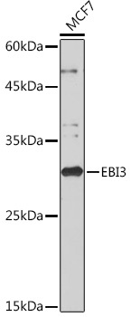
Western blot analysis of lysates from MCF7 cells, using EBI3 Rabbit mAb (CAB19613) at 1:1000 dilution. Secondary antibody: HRP-conjugated Goat anti-Rabbit IgG (H+L) (CABS014) at 1:10000 dilution. Lysates/proteins: 25μg per lane. Blocking buffer: 3% nonfat dry milk in TBST. Detection: ECL Basic Kit (AbGn00020). Exposure time: 10s.
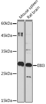
Western blot analysis of various lysates using EBI3 Rabbit mAb (CAB19613) at 1:1000 dilution. Secondary antibody: HRP-conjugated Goat anti-Rabbit IgG (H+L) (CABS014) at 1:10000 dilution. Lysates/proteins: 25μg per lane. Blocking buffer: 3% nonfat dry milk in TBST. Detection: ECL Basic Kit (AbGn00020). Exposure time: 90s.