The EED Antibody (CAB12773) is a high-quality antibody developed for reliable detection and analysis of target proteins. This antibody, produced in rabbits, shows high reactivity with human samples and is suitable for use in various applications including Western blot, immunofluorescence, and immunohistochemistry.EED is a key component of the Polycomb Repressive Complex 2 (PRC2), which plays a critical role in gene silencing and chromatin remodeling. Dysregulation of EED has been linked to various diseases including cancer, developmental disorders, and neurological conditions.
This antibody is validated for use in WB, IF/ICC, ELISA applications and has demonstrated reactivity against Human, Mouse, Rat samples.
Product Name:
EED Antibody
SKU:
CAB12773
Size:
20μL, 100μL
Reactivity:
Human, Mouse, Rat
Conjugate:
Unconjugated
Immunogen:
Synthetic peptide. This information is considered to be commercially sensitive.
Recommended starting concentration is 1 μg/mL. Please optimize the concentration based on your specific assay requirements.
Synonyms:
HEED, COGIS, WAIT1, EED
Positive Sample:
HeLa, MCF7, A-549, K-562, Mouse liver, Mouse lung, Rat lung
Cellular Localization:
Chromosome, Nucleus.
Calculated MW:
50kDa
Observed MW:
50-75kDa
This gene encodes a member of the Polycomb-group (PcG) family. PcG family members form multimeric protein complexes, which are involved in maintaining the transcriptional repressive state of genes over successive cell generations. This protein interacts with enhancer of zeste 2, the cytoplasmic tail of integrin beta7, immunodeficiency virus type 1 (HIV-1) MA protein, and histone deacetylase proteins. This protein mediates repression of gene activity through histone deacetylation, and may act as a specific regulator of integrin function. Two transcript variants encoding distinct isoforms have been identified for this gene.
Purification Method
Affinity purification
Gene ID
8726
RRID
AB_2759617
Buffer Information
Store at -20℃. Avoid freeze / thaw cycles. Buffer: PBS with 0.01% thimerosal,50% glycerol,pH7.3.
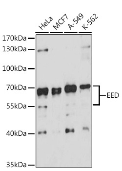
Western blot analysis of various lysates using EED Rabbit pAb (CAB12773) at 1:3000 dilution. Secondary antibody: HRP-conjugated Goat anti-Rabbit IgG (H+L) (CABS014) at 1:10000 dilution. Lysates/proteins: 25μg per lane. Blocking buffer: 3% nonfat dry milk in TBST. Detection: ECL Basic Kit (AbGn00020). Exposure time: 90s.
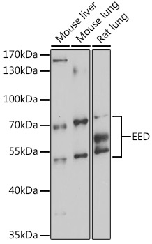
Western blot analysis of various lysates using EED Rabbit pAb (CAB12773) at 1:3000 dilution. Secondary antibody: HRP-conjugated Goat anti-Rabbit IgG (H+L) (CABS014) at 1:10000 dilution. Lysates/proteins: 25μg per lane. Blocking buffer: 3% nonfat dry milk in TBST. Detection: ECL Enhanced Kit (AbGn00021). Exposure time: 90s.
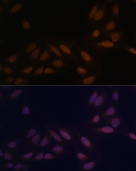
Immunofluorescence analysis of U-2 OS cells using EED Rabbit pAb (CAB12773) at dilution of 1:100 (40x lens). Secondary antibody: Cy3-conjugated Goat anti-Rabbit IgG (H+L) (CABS007) at 1:500 dilution. Blue: DAPI for nuclear staining.