The eIF4E Antibody (CAB2162) is a high-quality antibody developed for reliable detection and analysis of target proteins. The protein encoded by this gene is a component of the eukaryotic translation initiation factor 4F complex, which recognizes the 7-methylguanosine cap structure at the 5' end of messenger RNAs. The encoded protein aids in translation initiation by recruiting ribosomes to the 5'-cap structure. Association of this protein with the 4F complex is the rate-limiting step in translation initiation. This gene acts as a proto-oncogene, and its expression and activation is associated with transformation and tumorigenesis. Several pseudogenes of this gene are found on other chromosomes. Alternative splicing results in multiple transcript variants.
This antibody is validated for use in WB, IHC-P, IF/ICC, IP, ELISA applications and has demonstrated reactivity against Human, Mouse, Rat samples.
Product Name:
eIF4E Antibody
SKU:
CAB2162
Size:
100μL, 20μL
Reactivity:
Human, Mouse, Rat
Conjugate:
Unconjugated
Immunogen:
Synthetic peptide. This information is considered to be commercially sensitive.
Tested Applications:
WBIHC-PIF/ICCIPELISA
Recommended Dilution:
WB
1:500 - 1:1000
IHC-P
1:50 - 1:100
IF/ICC
1:50 - 1:200
IP
0.5μg-4μg antibody for 200μg-400μg extracts of whole cells
ELISA
Recommended starting concentration is 1 μg/mL. Please optimize the concentration based on your specific assay requirements.
Synonyms:
CBP, EIF4F, AUTS19, EIF4E1, eIF-4E, EIF4EL1, 4E
Positive Sample:
293T, HeLa, K-562
Cellular Localization:
Cytoplasm, P-Body.
Calculated MW:
25kDa
Observed MW:
25kDa/28kDa
The protein encoded by this gene is a component of the eukaryotic translation initiation factor 4F complex, which recognizes the 7-methylguanosine cap structure at the 5' end of messenger RNAs. The encoded protein aids in translation initiation by recruiting ribosomes to the 5'-cap structure. Association of this protein with the 4F complex is the rate-limiting step in translation initiation. This gene acts as a proto-oncogene, and its expression and activation is associated with transformation and tumorigenesis. Several pseudogenes of this gene are found on other chromosomes. Alternative splicing results in multiple transcript variants.
Purification Method
Affinity purification
Gene ID
1977
RRID
AB_2764180
Buffer Information
Store at -20℃. Avoid freeze / thaw cycles. Buffer: PBS containing 50% glycerol, preserved with proclin300 or sodium azide, pH 7.3.
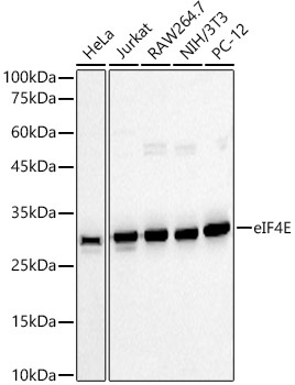
Western blot analysis of various lysates using eIF4E Rabbit pAb (CAB2162) at 1:1000 dilution. Secondary antibody: HRP-conjugated Goat anti-Rabbit IgG (H+L) (AS014) at 1:10000 dilution. Lysates/proteins: 25μg per lane. Blocking buffer: 3% nonfat dry milk in TBST. Detection: ECL Enhanced Kit (AbGn00021). Exposure time: 10s.
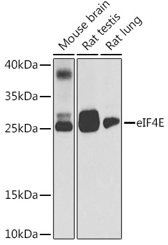
Western blot analysis of various lysates, using [KD Validated] eIF4E Rabbit pAb (CAB2162) at 1:500 dilution. Secondary antibody: HRP-conjugated Goat anti-Rabbit IgG (H+L) (AS014) at 1:10000 dilution. Lysates/proteins: 25μg per lane. Blocking buffer: 3% nonfat dry milk in TBST. Detection: ECL Basic Kit (AbGn00020). Exposure time: 30s.
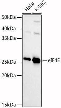
Western blot analysis of various lysates, using [KD Validated] eIF4E Rabbit pAb (CAB2162) at 1:1000 dilution. Secondary antibody: HRP-conjugated Goat anti-Rabbit IgG (H+L) (AS014) at 1:10000 dilution. Lysates/proteins: 25μg per lane. Blocking buffer: 3% nonfat dry milk in TBST. Detection: ECL Basic Kit (AbGn00020). Exposure time: 45s.
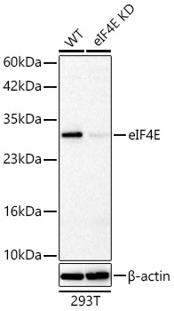
Western blot analysis of lysates from wild type (WT) and eIF4E knockdown (KD) 293T cells using [KD Validated] eIF4E Rabbit pAb (CAB2162) at 1:1400 dilution incubated overnight at 4℃. Secondary antibody: HRP-conjugated Goat anti-Rabbit IgG (H+L) (AS014) at 1:10000 dilution. Lysates/proteins: 25 μg per lane. Blocking buffer: 3% nonfat dry milk in TBST. Detection: ECL Basic Kit (AbGn00020) Exposure time: 10 s.
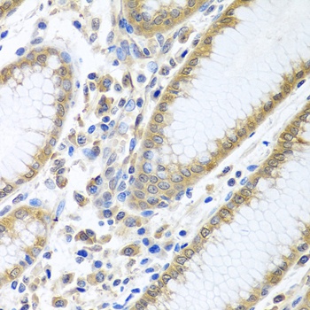
Immunohistochemistry analysis of paraffin-embedded Human stomach using [KD Validated] eIF4E Rabbit pAb (CAB2162) at dilution of 1:100 (40x lens). Microwave antigen retrieval performed with 0.01M PBS Buffer (pH 7.2) prior to IHC staining.
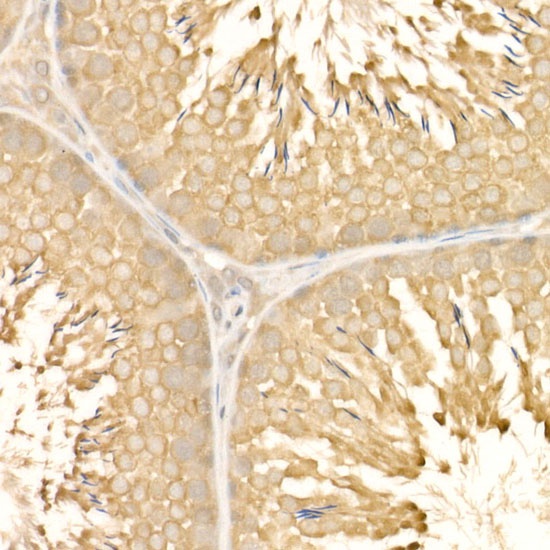
Immunohistochemistry analysis of paraffin-embedded Rat testis using [KD Validated] eIF4E Rabbit pAb (CAB2162) at dilution of 1:50 (40x lens). High pressure antigen retrieval performed with 0.01M Citrate buffer (pH 6.0) prior to IHC staining.
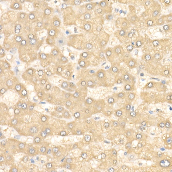
Immunohistochemistry analysis of paraffin-embedded Human liver using [KD Validated] eIF4E Rabbit pAb (CAB2162) at dilution of 1:50 (40x lens). High pressure antigen retrieval performed with 0.01M Citrate buffer (pH 6.0) prior to IHC staining.
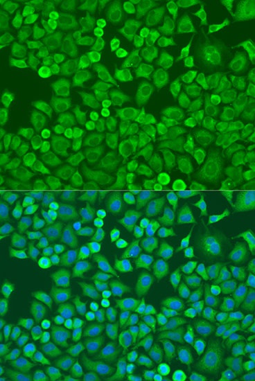
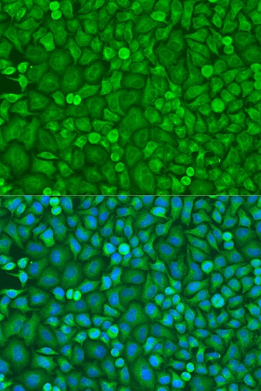
Immunofluorescence analysis of U2OS cells using eIF4E Rabbit pAb (CAB2162) at dilution of 1:100. Secondary antibody: Cy3-conjugated Goat anti-Rabbit IgG (H+L) (AS007) at 1:500 dilution. Blue: DAPI for nuclear staining.
Immunofluorescence analysis of U2OS cells using [KD Validated] eIF4E Rabbit pAb (CAB2162) at dilution of 1:100. Secondary antibody: Cy3-conjugated Goat anti-Rabbit IgG (H+L) (AS007) at 1:500 dilution. Blue: DAPI for nuclear staining.
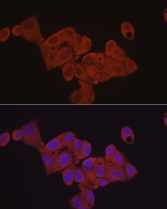
Immunofluorescence analysis of HepG2 cells using [KD Validated] eIF4E Rabbit pAb (CAB2162) at dilution of 1:50 (40x lens). Secondary antibody: Cy3-conjugated Goat anti-Rabbit IgG (H+L) (AS007) at 1:500 dilution. Blue: DAPI for nuclear staining.
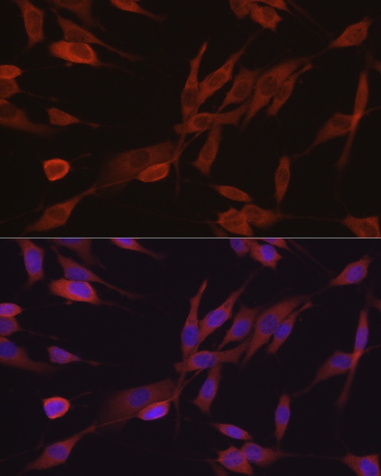
Immunofluorescence analysis of NIH/3T3 cells using [KD Validated] eIF4E Rabbit pAb (CAB2162) at dilution of 1:50 (40x lens). Secondary antibody: Cy3-conjugated Goat anti-Rabbit IgG (H+L) (AS007) at 1:500 dilution. Blue: DAPI for nuclear staining.
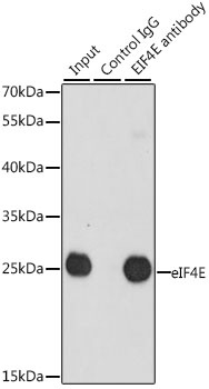
Immunoprecipitation analysis of 200 μg extracts of 293T cells, using 3 μg [KD Validated] eIF4E Rabbit pAb (CAB2162). Western blot was performed from the immunoprecipitate using [KD Validated] eIF4E Rabbit pAb at a dilution of 1:1000.