The EIF5 Antibody (CAB6583) is a high-quality antibody developed for reliable detection and analysis of target proteins. This antibody, produced in rabbits, is highly specific for EIF5 and has been validated for use in Western blot applications with human samples.EIF5 is involved in the assembly of the ribosomal preinitiation complex and plays a crucial role in the regulation of translation initiation. Dysregulation of EIF5 has been linked to various diseases, including cancer and neurodegenerative disorders, making it an important target for research in molecular biology and translational medicine.
This antibody is validated for use in WB, IF/ICC, ELISA applications and has demonstrated reactivity against Human, Mouse, Rat samples.
Product Name:
EIF5 Antibody
SKU:
CAB6583
Size:
20μL, 100μL
Reactivity:
Human, Mouse, Rat
Conjugate:
Unconjugated
Immunogen:
Recombinant protein (or fragment).This information is considered to be commercially sensitive.
Recommended starting concentration is 1 μg/mL. Please optimize the concentration based on your specific assay requirements.
Synonyms:
EIF-5, EIF-5A, EIF5
Positive Sample:
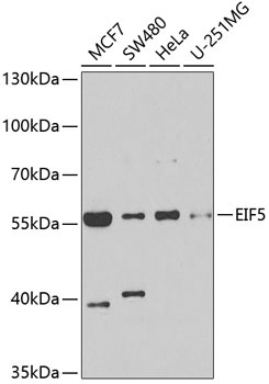
MCF7, SW480, HeLa, U-251MG
Cellular Localization:
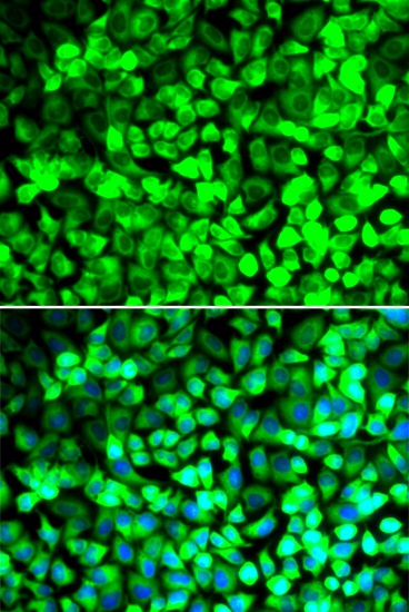
Cytoplasm, Cytosol, Plasma Membrane, Synapse.
Calculated MW:
49kDa
Observed MW:
55kDa
Eukaryotic translation initiation factor-5 (EIF5) interacts with the 40S initiation complex to promote hydrolysis of bound GTP with concomitant joining of the 60S ribosomal subunit to the 40S initiation complex. The resulting functional 80S ribosomal initiation complex is then active in peptidyl transfer and chain elongations (summary by Si et al., 1996 [PubMed 8663286]).
Purification Method
Affinity purification
Gene ID
1983
RRID
AB_2767176
Buffer Information
Store at -20℃. Avoid freeze / thaw cycles. Buffer: PBS containing 50% glycerol, preserved with proclin300 or sodium azide, pH 7.3.
Western blot analysis of various lysates using EIF5 Rabbit pAb (CAB6583) at 1:1000 dilution. Secondary antibody: HRP-conjugated Goat anti-Rabbit IgG (H+L) (CABS014) at 1:10000 dilution. Lysates/proteins: 25μg per lane. Blocking buffer: 3% nonfat dry milk in TBST. Detection: ECL Basic Kit (AbGn00020). Exposure time: 90s.
Immunofluorescence analysis of U2OS cells using EIF5 Rabbit pAb (CAB6583). Secondary antibody: Cy3-conjugated Goat anti-Rabbit IgG (H+L) (CABS007) at 1:500 dilution. Blue: DAPI for nuclear staining.