The EIF5B Antibody (CAB15123) is a high-quality antibody developed for reliable detection and analysis of target proteins. This antibody, produced in rabbits, displays high specificity and sensitivity towards human samples, making it a reliable choice for Western blot applications. By binding to EIF5B, it enables researchers to detect and analyze the protein in various cell types, facilitating studies in molecular biology and cancer research.EIF5B is a critical component of the translation initiation complex, playing a crucial role in the accurate start of protein synthesis. Dysregulation of EIF5B has been linked to various diseases, including cancer, making it an intriguing target for therapeutic intervention.
This antibody is validated for use in WB, IF/ICC, ELISA applications and has demonstrated reactivity against Human, Mouse, Rat samples.
Product Name:
EIF5B Antibody
SKU:
CAB15123
Size:
20μL, 100μL
Reactivity:
Human, Mouse, Rat
Conjugate:
Unconjugated
Immunogen:
Recombinant protein (or fragment).This information is considered to be commercially sensitive.
Recommended starting concentration is 1 μg/mL. Please optimize the concentration based on your specific assay requirements.
Synonyms:
IF2, EIF5B
Positive Sample:
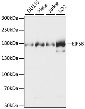
DU145, HeLa, Jurkat, LO2
Cellular Localization:
Cytoplasm.
Calculated MW:
139kDa
Observed MW:
179kDa
Accurate initiation of translation in eukaryotes is complex and requires many factors, some of which are composed of multiple subunits. The process is simpler in prokaryotes which have only three initiation factors (IF1, IF2, IF3). Two of these factors are conserved in eukaryotes: the homolog of IF1 is eIF1A and the homolog of IF2 is eIF5B. This gene encodes eIF5B. Factors eIF1A and eIF5B interact on the ribosome along with other initiation factors and GTP to position the initiation methionine tRNA on the start codon of the mRNA so that translation initiates accurately.
Purification Method
Affinity purification
Gene ID
9669
RRID
AB_2762008
Buffer Information
Store at -20℃. Avoid freeze / thaw cycles. Buffer: PBS with 0.01% thimerosal,50% glycerol,pH7.3.
Western blot analysis of various lysates using EIF5B Rabbit pAb (CAB15123). Secondary antibody: HRP-conjugated Goat anti-Rabbit IgG (H+L) (CABS014) at 1:10000 dilution. Lysates/proteins: 25μg per lane. Blocking buffer: 3% nonfat dry milk in TBST. Detection: ECL Basic Kit (AbGn00020). Exposure time: 3s.
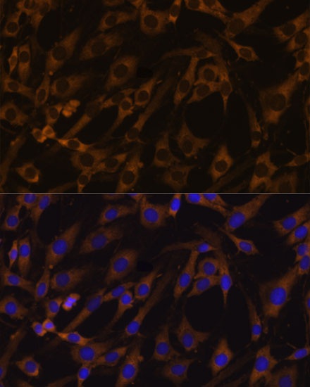
Immunofluorescence analysis of C6 cells using EIF5B Rabbit pAb (CAB15123) at dilution of 1:100. Secondary antibody: Cy3-conjugated Goat anti-Rabbit IgG (H+L) (CABS007) at 1:500 dilution. Blue: DAPI for nuclear staining.
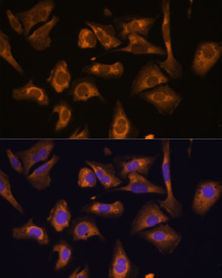
Immunofluorescence analysis of U-2 OS cells using EIF5B Rabbit pAb (CAB15123) at dilution of 1:100. Secondary antibody: Cy3-conjugated Goat anti-Rabbit IgG (H+L) (CABS007) at 1:500 dilution. Blue: DAPI for nuclear staining.