The ErbB4/HER4 Antibody (CAB6133) is a high-quality antibody developed for reliable detection and analysis of target proteins. This antibody, produced in rabbits, exhibits high reactivity with human samples and is suitable for use in Western blot applications. By binding specifically to the ERBB4 protein, researchers can accurately detect and analyze its expression levels in a variety of cell types.ERBB4, also known as HER4, is a crucial player in signal transduction pathways that regulate cell proliferation and survival.
This antibody is validated for use in WB, IHC-P, IF/ICC, ELISA applications and has demonstrated reactivity against Human, Mouse, Rat samples.
Product Name:
ErbB4/HER4 Antibody
SKU:
CAB6133
Size:
20μL, 100μL
Reactivity:
Human, Mouse, Rat
Conjugate:
Unconjugated
Immunogen:
Synthetic peptide. This information is considered to be commercially sensitive.
Recommended starting concentration is 1 μg/mL. Please optimize the concentration based on your specific assay requirements.
Synonyms:
HER4, ALS19, p180erbB4, ErbB4/HER4
Positive Sample:
Mouse brain
Cellular Localization:
Cell Membrane, Mitochondrion, Nucleus, Single-Pass Type I Membrane Protein.
Calculated MW:
147kDa
Observed MW:
180kDa
This gene is a member of the Tyr protein kinase family and the epidermal growth factor receptor subfamily. It encodes a single-pass type I membrane protein with multiple cysteine rich domains, a transmembrane domain, a tyrosine kinase domain, a phosphotidylinositol-3 kinase binding site and a PDZ domain binding motif. The protein binds to and is activated by neuregulins and other factors and induces a variety of cellular responses including mitogenesis and differentiation. Multiple proteolytic events allow for the release of a cytoplasmic fragment and an extracellular fragment. Mutations in this gene have been associated with cancer. Alternatively spliced variants which encode different protein isoforms have been described; however, not all variants have been fully characterized.
Purification Method
Affinity purification
Gene ID
2066
RRID
AB_2766764
Buffer Information
Store at -20℃. Avoid freeze / thaw cycles. Buffer: PBS with 0.09% Sodium azide,50% glycerol,pH7.3.
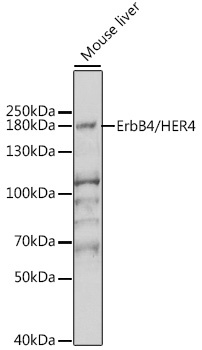
Western blot analysis of lysates from mouse brain, using ErbB4/HER4 Rabbit pAb (CAB6133) at 1:1000 dilution. Secondary antibody: HRP-conjugated Goat anti-Rabbit IgG (H+L) (CABS014) at 1:10000 dilution. Lysates/proteins: 25μg per lane. Blocking buffer: 3% nonfat dry milk in TBST. Detection: ECL Enhanced Kit (AbGn00021). Exposure time: 10s.
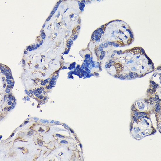
Immunohistochemistry analysis of paraffin-embedded Human placenta using ErbB4/HER4 Rabbit pAb (CAB6133) at dilution of 1:100 (40x lens). High pressure antigen retrieval performed with 0.01M Citrate buffer (pH 6.0) prior to IHC staining.
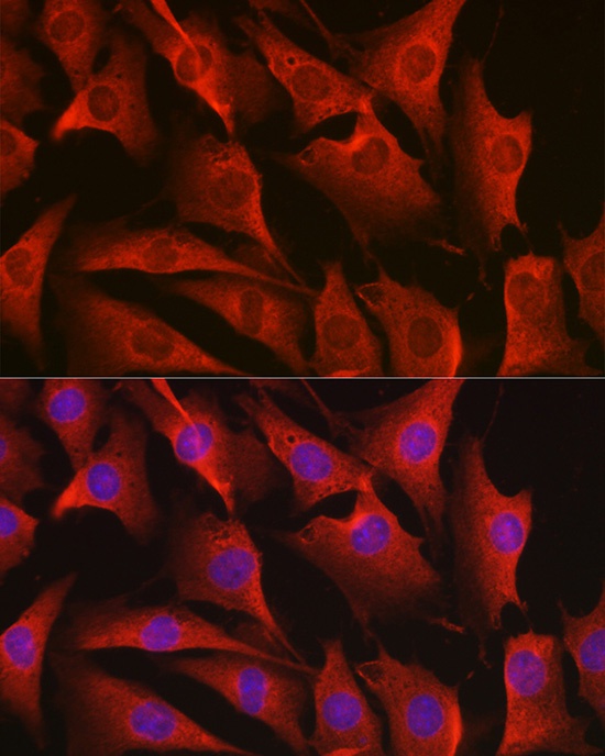
Immunofluorescence analysis of NIH-3T3 cells using ErbB4/HER4 Rabbit pAb (CAB6133) at dilution of 1:100 (40x lens). Secondary antibody: Cy3-conjugated Goat anti-Rabbit IgG (H+L) (CABS007) at 1:500 dilution. Blue: DAPI for nuclear staining.
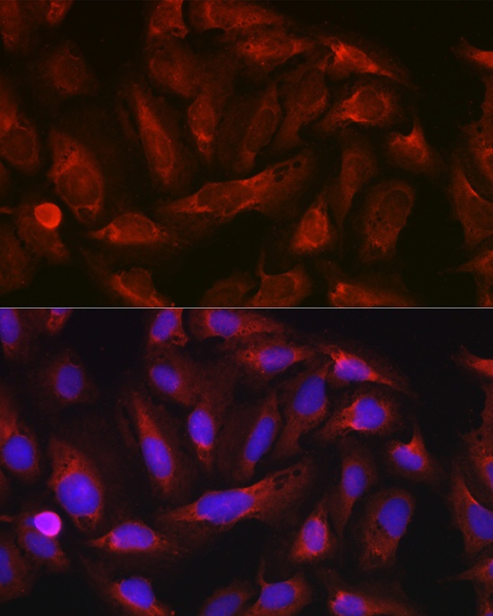
Immunofluorescence analysis of U-2 OS cells using ErbB4/HER4 Rabbit pAb (CAB6133) at dilution of 1:100 (40x lens). Secondary antibody: Cy3-conjugated Goat anti-Rabbit IgG (H+L) (CABS007) at 1:500 dilution. Blue: DAPI for nuclear staining.


")
 (Active) (RPES3837)")