The ERK1 Monoclonal Antibody (CAB19561) is a high-quality antibody developed for reliable detection and analysis of target proteins. ERK1 is a key regulator of cellular proliferation, differentiation, and survival, making it a crucial target for studies in cell signaling and cancer research.This antibody, produced in rabbits, exhibits high reactivity with human samples and has been validated for use in Western blot applications. It specifically binds to the ERK1 protein, enabling precise detection and analysis in various cell types. This makes it an ideal tool for researchers interested in understanding the role of ERK1 in normal cellular processes and disease pathways.
This antibody is validated for use in WB, IHC-P, IF/ICC, IP, ELISA, IF-P applications and has demonstrated reactivity against Human, Mouse samples.
Product Name:
ERK1 Monoclonal Antibody
SKU:
CAB19561
Size:
20μL, 100μL
Reactivity:
Human, Mouse
Clone Number:
ARC2591
Conjugate:
Unconjugated
Immunogen:
Synthetic peptide. This information is considered to be commercially sensitive.
The protein encoded by this gene is a member of the MAP kinase family. MAP kinases, also known as extracellular signal-regulated kinases (ERKs), act in a signaling cascade that regulates various cellular processes such as proliferation, differentiation, and cell cycle progression in response to a variety of extracellular signals. This kinase is activated by upstream kinases, resulting in its translocation to the nucleus where it phosphorylates nuclear targets. Alternatively spliced transcript variants encoding different protein isoforms have been described.
Purification Method
Affinity purification
Gene ID
5595
RRID
AB_2862668
Buffer Information
Store at -20℃. Avoid freeze / thaw cycles. Buffer: PBS containing 50% glycerol and 0.05% BSA, preserved with proclin300 or sodium azide, pH 7.3.
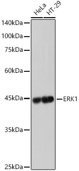
Western blot analysis of various lysates, using [KD Validated] ERK1 Rabbit mAb (CAB19561) at 1:1000 dilution. Secondary antibody: HRP-conjugated Goat anti-Rabbit IgG (H+L) (CABS014) at 1:10000 dilution. Lysates/proteins: 25μg per lane. Blocking buffer: 3% nonfat dry milk in TBST. Detection: ECL Basic Kit (AbGn00020). Exposure time: 1s.
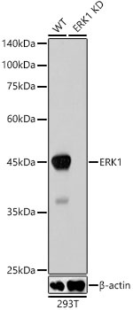
Western blot analysis of lysates from wild type(WT) and ERK1 knockdown (KD) 293T cells, using [KD Validated] ERK1 Rabbit mAb (CAB19561) at 1:1000 dilution. Secondary antibody: HRP-conjugated Goat anti-Rabbit IgG (H+L) (CABS014) at 1:10000 dilution. Lysates/proteins: 25μg per lane. Blocking buffer: 3% nonfat dry milk in TBST. Detection: ECL Basic Kit (AbGn00020). Exposure time: 1s.
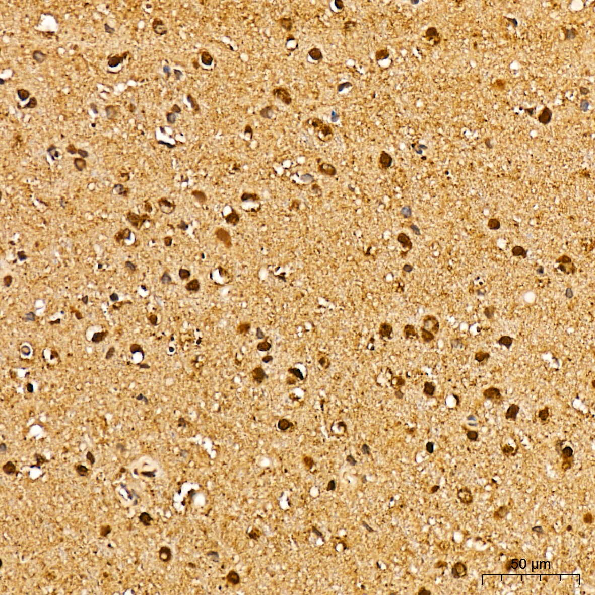
Immunohistochemistry analysis of paraffin-embedded Human brain tissue using [KD Validated] ERK1 Rabbit mAb (CAB19561) at a dilution of 1:200 (40x lens). High pressure antigen retrieval was performed with 0.01 M citrate buffer (pH 6.0) prior to IHC staining.
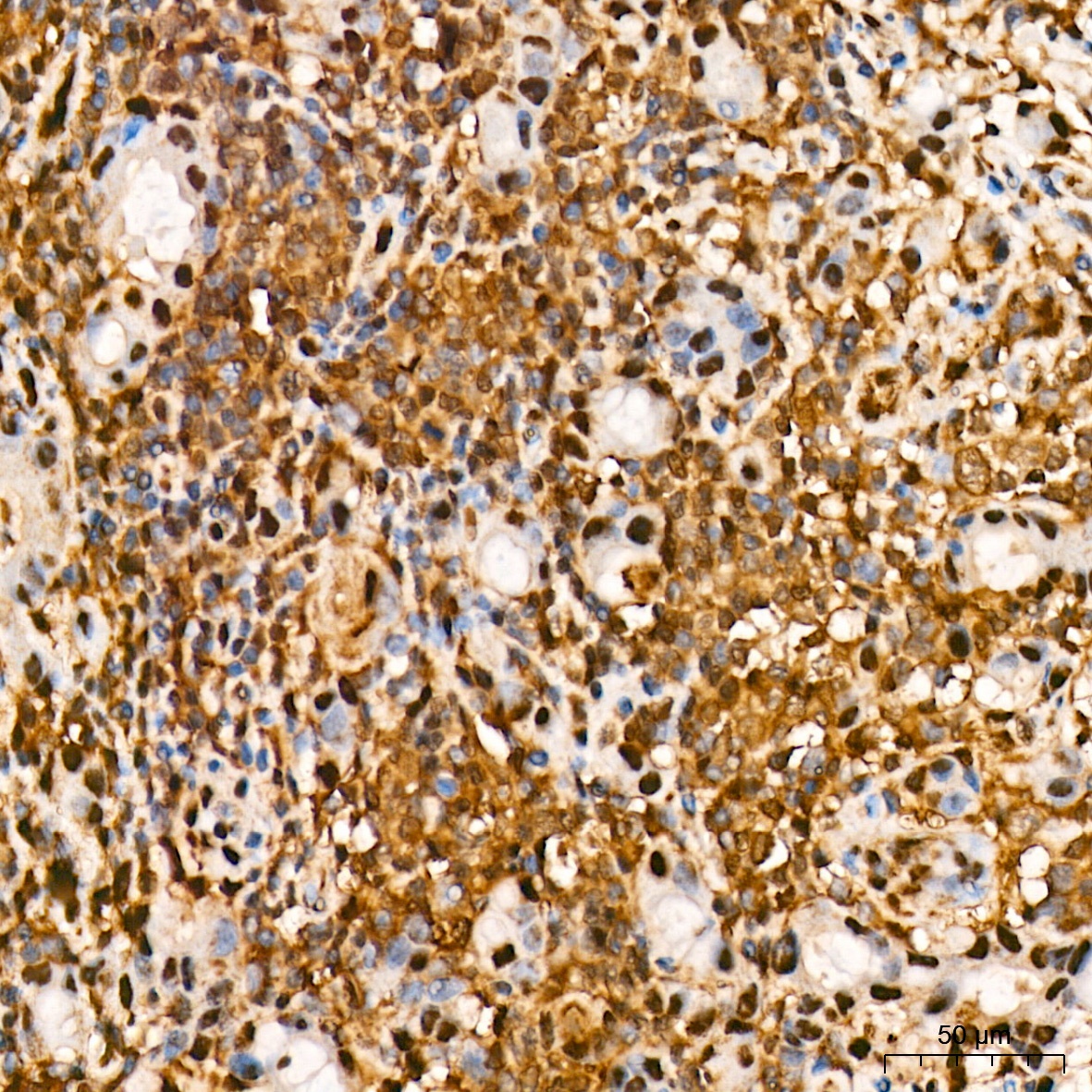
Immunohistochemistry analysis of paraffin-embedded Human breast tissue using [KD Validated] ERK1 Rabbit mAb (CAB19561) at a dilution of 1:200 (40x lens). High pressure antigen retrieval was performed with 0.01 M citrate buffer (pH 6.0) prior to IHC staining.
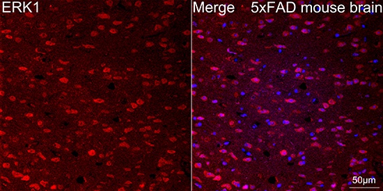
Confocal imaging of paraffin-embedded 5xFAD mouse brain tissue using [KD Validated] ERK1 Rabbit mAb (CAB19561, dilution 1:100) followed by a further incubation with Cy3 Goat Anti-Rabbit IgG (H+L) (CABS007, dilution 1:500) (Red). DAPI was used for nuclear staining (Blue). Objective: 40x. Perform microwave antigen retrieval with 0.01 M citrate buffer (pH 6.0) prior to IF staining.
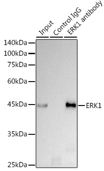
Immunoprecipitation analysis of 300 μg extracts of HeLa cells using 3 μg [KD Validated] ERK1 Rabbit mAb (CAB19561). Western blot was performed from the immunoprecipitate using ERK1 antibody (CAB19561) at a dilution of 1:1000.