The F4/80 Monoclonal Antibody (CAB23788) is a high-quality antibody developed for reliable detection and analysis of target proteins. This antibody, produced using hybridoma technology, specifically targets the F4/80 protein found on the surface of macrophages in mouse samples. With a high level of specificity and sensitivity, the F4/80 Monoclonal Antibody is suitable for use in various applications including immunohistochemistry, flow cytometry, and immunofluorescence. Its ability to bind to the F4/80 protein allows for precise detection and analysis of macrophages in different tissues and biological samples.
This antibody is validated for use in IHC-P, ELISA, IF-P applications and has demonstrated reactivity against Mouse, Rat samples.
Product Name:
F4/80 Monoclonal Antibody
SKU:
CAB23788
Size:
20μL, 100μL
Reactivity:
Mouse, Rat
Clone Number:
ARC61555
Conjugate:
Unconjugated
Immunogen:
Recombinant protein (or fragment).This information is considered to be commercially sensitive.
Predicted to enable G protein-coupled receptor activity. Predicted to be involved in adenylate cyclase-activating G protein-coupled receptor signaling pathway. Predicted to act upstream of or within G protein-coupled receptor signaling pathway and adaptive immune response. Located in external side of plasma membrane. Is expressed in several structures, including cardiovascular system; central nervous system; genitourinary system; hemolymphoid system; and intestine. Orthologous to human ADGRE1 (adhesion G protein-coupled receptor E1).
Purification Method
Affinity purification
Gene ID
13733
Buffer Information
Store at -20℃. Avoid freeze / thaw cycles. Buffer: PBS containing 50% glycerol and 0.05% BSA, preserved with proclin300 or sodium azide, pH 7.3.
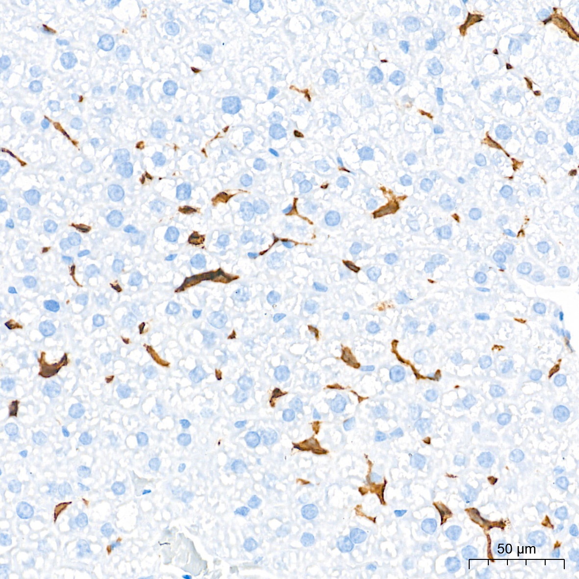
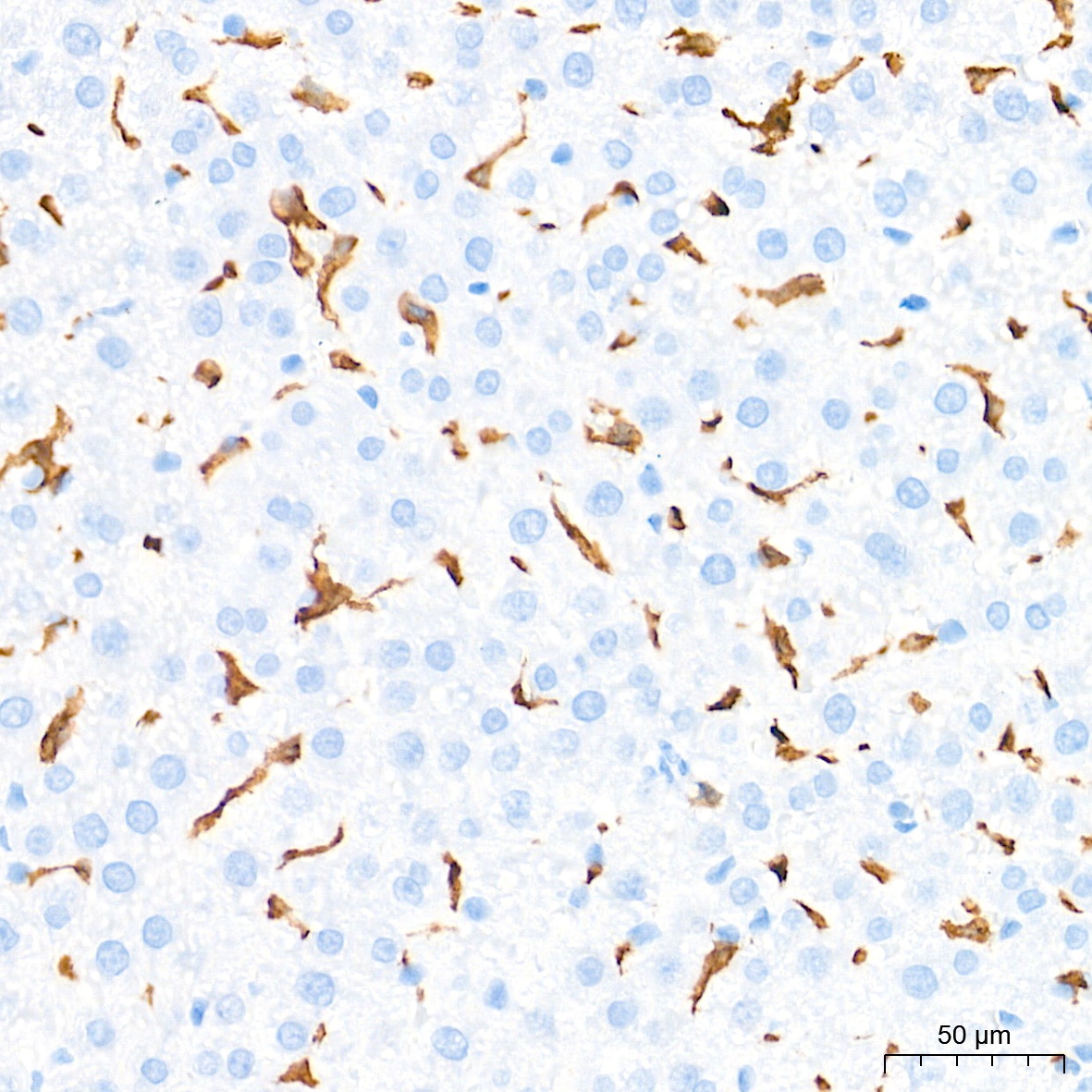
Immunohistochemistry analysis of paraffin-embedded Mouse liver tissue using F4/80 Rabbit mAb (CAB23788) at a dilution of 1:500 (40x lens). High pressure antigen retrieval performed with 0.01M Tris-EDTA Buffer (pH 9.0) prior to IHC staining.
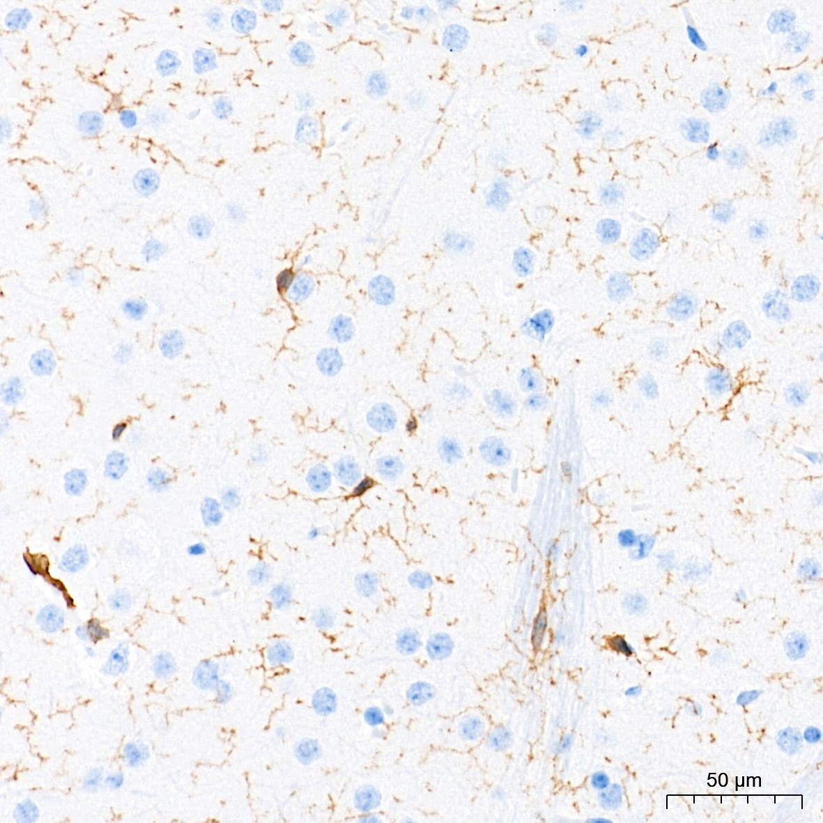
Immunohistochemistry analysis of paraffin-embedded Mouse brain tissue using F4/80 Rabbit mAb (CAB23788) at a dilution of 1:500 (40x lens). High pressure antigen retrieval performed with 0.01M Tris-EDTA Buffer (pH 9.0) prior to IHC staining.
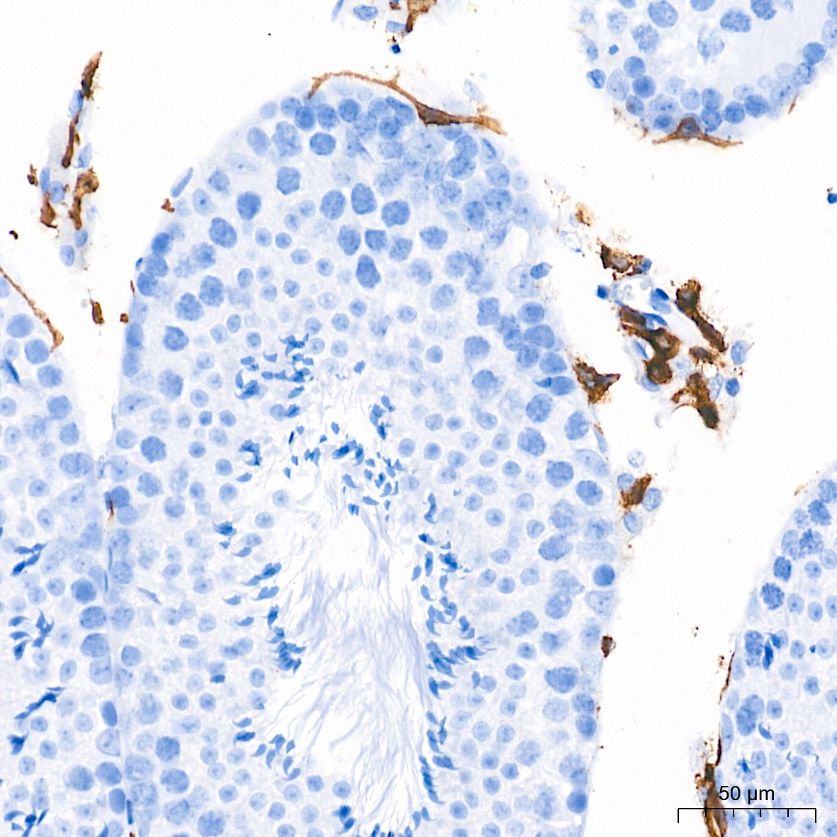
Immunohistochemistry analysis of paraffin-embedded Mouse testis tissue using F4/80 Rabbit mAb (CAB23788) at a dilution of 1:500 (40x lens). High pressure antigen retrieval performed with 0.01M Tris-EDTA Buffer (pH 9.0) prior to IHC staining.
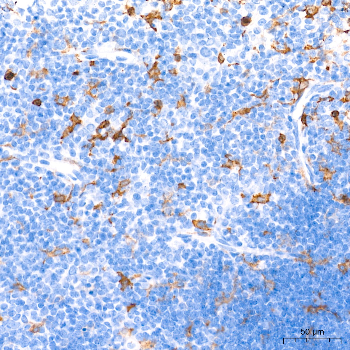
Immunohistochemistry analysis of paraffin-embedded Mouse thymus tissue using F4/80 Rabbit mAb (CAB23788) at a dilution of 1:500 (40x lens). High pressure antigen retrieval performed with 0.01M Tris-EDTA Buffer (pH 9.0) prior to IHC staining.
Immunohistochemistry analysis of paraffin-embedded Rat liver tissue using F4/80 Rabbit mAb (CAB23788) at a dilution of 1:500 (40x lens). High pressure antigen retrieval performed with 0.01M Tris-EDTA Buffer (pH 9.0) prior to IHC staining.
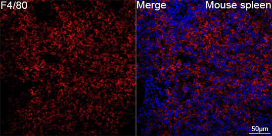
Confocal imaging of paraffin-embedded Mouse spleen tissue using F4/80 Rabbit mAb (CAB23788, dilution 1:200) followed by a further incubation with Cy3 Goat Anti-Rabbit IgG (H+L) (CABS007, dilution 1:500) (Red). DAPI was used for nuclear staining (Blue). High pressure antigen retrieval performed with 0.01M Citrate Buffer (pH 6.0) prior to IF staining. Objective: 40x.
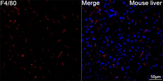
Confocal imaging of paraffin-embedded Mouse liver tissue using F4/80 Rabbit mAb (CAB23788, dilution 1:200) followed by a further incubation with Cy3 Goat Anti-Rabbit IgG (H+L) (CABS007, dilution 1:500) (Red). DAPI was used for nuclear staining (Blue). High pressure antigen retrieval performed with 0.01M Citrate Buffer (pH 6.0) prior to IF staining. Objective: 40x.
 at 1:1000 dilution. Secondary antibody: HRP Goat Anti-Rabbit IgG (H+L) at 1:10000 dilution. Lysates/proteins: 25μg per lane. Blocking buffer: 3% nonfat dry milk in TBST.")
 at 1:1000 dilution. Secondary antibody: HRP Goat Anti-Rabbit IgG (H+L) at 1:10000 dilution. Lysates/proteins: 25μg per lane. Blocking buffer: 3% nonfat dry milk in TBST.")

![Biotin Anti-Mouse F4/80 Antibody [CI:A3-1] (AGEL0043)](https://cdn11.bigcommerce.com/s-h68l9z2lnx/images/stencil/590x590/products/20014/605915/biotin-anti-mouse-f480-antibody-cia3-1-agel0043__83745.1707496384.jpg?c=2 "Biotin Anti-Mouse F4/80 Antibody [CI:A3-1] (AGEL0043)")
![APC Anti-Mouse F4/80 Antibody [CI:A3-1] (AGEL0568)](https://cdn11.bigcommerce.com/s-h68l9z2lnx/images/stencil/590x590/products/21099/606223/apc-anti-mouse-f480-antibody-cia3-1-agel0568__95503.1707497344.jpg?c=2 "APC Anti-Mouse F4/80 Antibody [CI:A3-1] (AGEL0568)")
![APC Anti-Mouse F4/80 Antibody [CI:A3-1] (AGEL0568)](https://cdn11.bigcommerce.com/s-h68l9z2lnx/images/stencil/590x590/products/21099/719577/AGEL0568_spectral__88907.1775209664.png?c=2 "APC Anti-Mouse F4/80 Antibody [CI:A3-1] (AGEL0568)")
![FITC Anti-Mouse F4/80 Antibody [CI:A3-1] (AGEL0566)](https://cdn11.bigcommerce.com/s-h68l9z2lnx/images/stencil/590x590/products/237378/618661/fitc-anti-mouse-f480-antibody-cia3-1-agel0566__33820.1733541893.jpg?c=2 "FITC Anti-Mouse F4/80 Antibody [CI:A3-1] (AGEL0566)")
![FITC Anti-Mouse F4/80 Antibody [CI:A3-1] (AGEL0566)](https://cdn11.bigcommerce.com/s-h68l9z2lnx/images/stencil/590x590/products/237378/719575/AGEL0566_spectral__43731.1775209660.png?c=2 "FITC Anti-Mouse F4/80 Antibody [CI:A3-1] (AGEL0566)")
![PE Anti-Mouse F4/80 Antibody [CI:A3-1] (AGEL0567)](https://cdn11.bigcommerce.com/s-h68l9z2lnx/images/stencil/590x590/products/21098/606435/pe-anti-mouse-f480-antibody-cia3-1-agel0567__88252.1707498046.jpg?c=2 "PE Anti-Mouse F4/80 Antibody [CI:A3-1] (AGEL0567)")
![PE Anti-Mouse F4/80 Antibody [CI:A3-1] (AGEL0567)](https://cdn11.bigcommerce.com/s-h68l9z2lnx/images/stencil/590x590/products/21098/719576/AGEL0567_spectral__61893.1775209662.png?c=2 "PE Anti-Mouse F4/80 Antibody [CI:A3-1] (AGEL0567)")