The FCRL3 Antibody (CAB10452) is a high-quality antibody developed for reliable detection and analysis of target proteins. This antibody, produced in rabbits, is highly specific for FCRL3 in human samples and has been validated for use in Western blot applications.FCRL3 is known to be involved in regulating immune responses, making it a promising target for research in immunology and cancer biology. By detecting and analyzing FCRL3 protein expression in various cell types, this antibody enables researchers to gain insights into the function of FCRL3 in different disease contexts.
This antibody is validated for use in WB, ELISA applications and has demonstrated reactivity against Human, Mouse samples.
Product Name:
FCRL3 Antibody
SKU:
CAB10452
Size:
20μL, 100μL
Reactivity:
Human, Mouse
Conjugate:
Unconjugated
Immunogen:
Recombinant protein (or fragment).This information is considered to be commercially sensitive.
Recommended starting concentration is 1 μg/mL. Please optimize the concentration based on your specific assay requirements.
Synonyms:
MAIA, FCRH3, IFGP3, IRTA3, SPAP2, CD307c, FCRL3
Positive Sample:
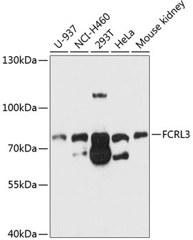
U-937, NCI-H460, 293T, HeLa, Mouse kidney
Cellular Localization:
Cell Membrane, Single-Pass Type I Membrane Protein.
Calculated MW:
81kDa
Observed MW:
81kDa
This gene encodes a member of the immunoglobulin receptor superfamily and is one of several Fc receptor-like glycoproteins clustered on the long arm of chromosome 1. The encoded protein contains immunoreceptor-tyrosine activation motifs and immunoreceptor-tyrosine inhibitory motifs in its cytoplasmic domain and may play a role in regulation of the immune system. Mutations in this gene have been associated with rheumatoid arthritis, autoimmune thyroid disease, and systemic lupus erythematosus. Alternative splicing results in multiple transcript variants.
Purification Method
Affinity purification
Gene ID
115352
RRID
AB_2758000
Buffer Information
Store at -20℃. Avoid freeze / thaw cycles. Buffer: PBS containing 50% glycerol, preserved with proclin300 or sodium azide, pH 7.3.
Western blot analysis of various lysates using FCRL3 Rabbit pAb (CAB10452) at 1:1000 dilution. Secondary antibody: HRP-conjugated Goat anti-Rabbit IgG (H+L) (CABS014) at 1:10000 dilution. Lysates/proteins: 25μg per lane. Blocking buffer: 3% nonfat dry milk in TBST. Detection: ECL Basic Kit (AbGn00020). Exposure time: 30s.