The FEN-1 Monoclonal Antibody (CAB8999) is a high-quality antibody developed for reliable detection and analysis of target proteins. This antibody, raised in rabbits, is highly reactive with human samples and is validated for use in Western blot applications. It binds specifically to the FEN1 protein, allowing for accurate detection and analysis in various cell types, making it ideal for studies in genetics, cancer research, and other areas of molecular biology.FEN1, also known as flap endonuclease 1, is essential for maintaining genomic stability by processing DNA intermediates during DNA replication and repair.
This antibody is validated for use in WB, IHC-P, ELISA applications and has demonstrated reactivity against Human, Mouse samples.
Product Name:
FEN-1 Monoclonal Antibody
SKU:
CAB8999
Size:
20μL, 100μL
Reactivity:
Human, Mouse
Clone Number:
ARC1376
Conjugate:
Unconjugated
Immunogen:
Synthetic peptide. This information is considered to be commercially sensitive.
Recommended starting concentration is 1 μg/mL. Please optimize the concentration based on your specific assay requirements.
Synonyms:
MF1, RAD2, FEN-1
Positive Sample:
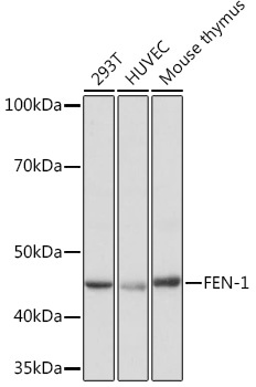
293T, HUVEC, Mouse thymus
Cellular Localization:
Mitochondrion, Nucleus, Nucleolus, Nucleoplasm.
Calculated MW:
43kDa
Observed MW:
45kDa
The protein encoded by this gene removes 5' overhanging flaps in DNA repair and processes the 5' ends of Okazaki fragments in lagging strand DNA synthesis. Direct physical interaction between this protein and AP endonuclease 1 during long-patch base excision repair provides coordinated loading of the proteins onto the substrate, thus passing the substrate from one enzyme to another. The protein is a member of the XPG/RAD2 endonuclease family and is one of ten proteins essential for cell-free DNA replication. DNA secondary structure can inhibit flap processing at certain trinucleotide repeats in a length-dependent manner by concealing the 5' end of the flap that is necessary for both binding and cleavage by the protein encoded by this gene. Therefore, secondary structure can deter the protective function of this protein, leading to site-specific trinucleotide expansions.
Purification Method
Affinity purification
Gene ID
2237
RRID
AB_2863639
Buffer Information
Store at -20℃. Avoid freeze / thaw cycles. Buffer: PBS containing 50% glycerol and 0.05% BSA, preserved with proclin300 or sodium azide, pH 7.3.
Western blot analysis of various lysates using FEN-1 Rabbit mAb (CAB8999) at 1:1000 dilution. Secondary antibody: HRP-conjugated Goat anti-Rabbit IgG (H+L) (CABS014) at 1:10000 dilution. Lysates/proteins: 25μg per lane. Blocking buffer: 3% nonfat dry milk in TBST. Detection: ECL Basic Kit (AbGn00020). Exposure time: 1s.
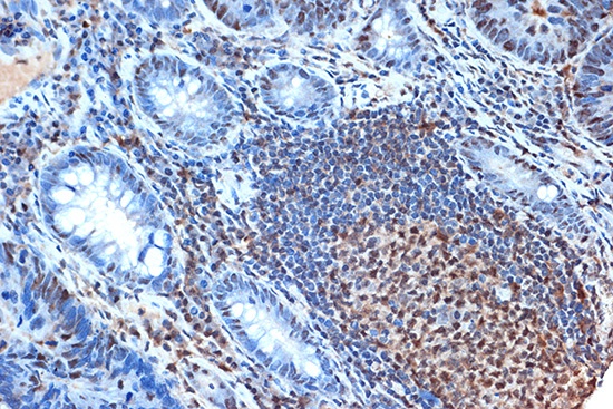
Immunohistochemistry analysis of paraffin-embedded Human colon carcinoma using FEN-1 Rabbit mAb (CAB8999) at dilution of 1:100 (40x lens). Microwave antigen retrieval performed with 0.01M Tris/EDTA Buffer (pH 9.0) prior to IHC staining.