Ferritin Heavy Chain Monoclonal Antibody (CAB19544)
The Ferritin Heavy Chain Monoclonal Antibody (CAB19544) is a high-quality antibody developed for reliable detection and analysis of target proteins. This gene encodes the heavy subunit of ferritin, the major intracellular iron storage protein in prokaryotes and eukaryotes. It is composed of 24 subunits of the heavy and light ferritin chains. Variation in ferritin subunit composition may affect the rates of iron uptake and release in different tissues. A major function of ferritin is the storage of iron in a soluble and nontoxic state. Defects in ferritin proteins are associated with several neurodegenerative diseases. This gene has multiple pseudogenes. Several alternatively spliced transcript variants have been observed, but their biological validity has not been determined.
This antibody is validated for use in WB, ELISA, IF-P applications and has demonstrated reactivity against Human, Mouse, Rat samples.
Product Name:
Ferritin Heavy Chain Monoclonal Antibody
SKU:
CAB19544
Size:
100μL, 20μL
Reactivity:
Human, Mouse, Rat
Clone Number:
ARC0023
Conjugate:
Unconjugated
Immunogen:
Synthetic peptide. This information is considered to be commercially sensitive.
Tested Applications:
WBELISAIF-P
Recommended Dilution:
WB
1:1000 - 1:10000
IF-P
1:50 - 1:200
ELISA
Recommended starting concentration is 1 μg/mL. Please optimize the concentration based on your specific assay requirements.
Synonyms:
FHC, FTH, HFE5, PLIF, FTHL6, PIG15, Ferritin Heavy Chain
This gene encodes the heavy subunit of ferritin, the major intracellular iron storage protein in prokaryotes and eukaryotes. It is composed of 24 subunits of the heavy and light ferritin chains. Variation in ferritin subunit composition may affect the rates of iron uptake and release in different tissues. A major function of ferritin is the storage of iron in a soluble and nontoxic state. Defects in ferritin proteins are associated with several neurodegenerative diseases. This gene has multiple pseudogenes. Several alternatively spliced transcript variants have been observed, but their biological validity has not been determined.
Purification Method
Affinity purification
Gene ID
2495
RRID
AB_2862659
Buffer Information
Store at -20℃. Avoid freeze / thaw cycles. Buffer: PBS containing 50% glycerol and 0.05% BSA, preserved with proclin300 or sodium azide, pH 7.3.
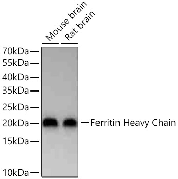
Western blot analysis of various lysates using Ferritin Heavy Chain Rabbit mAb (CAB19544) at 1:2000 dilution incubated overnight at 4℃. Secondary antibody: HRP-conjugated Goat anti-Rabbit IgG (H+L) (AS014) at 1:10000 dilution. Lysates/proteins: 25 μg per lane. Blocking buffer: 3% nonfat dry milk in TBST. Detection: ECL Basic Kit (AbGn00020). Exposure time: 1s.
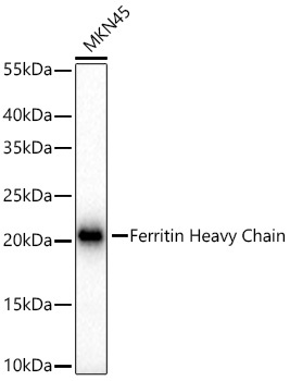
Western blot analysis of lysates from MKN45 cells using Ferritin Heavy Chain Rabbit mAb (CAB19544) at 1:1000 dilution incubated overnight at 4℃. Secondary antibody: HRP-conjugated Goat anti-Rabbit IgG (H+L) (AS014) at 1:10000 dilution. Lysates/proteins: 25 μg per lane. Blocking buffer: 3% nonfat dry milk in TBST. Detection: ECL Basic Kit (AbGn00020). Exposure time: 1s.
Confocal imaging of paraffin-embedded Mouse liver tissue using Ferritin Heavy Chain Rabbit mAb (CAB19544, dilution 1:200) followed by a further incubation with Cy3 Goat Anti-Rabbit IgG (H+L) (AS007, dilution 1:500) (Red). DAPI was used for nuclear staining (Blue). High pressure antigen retrieval performed with 0.01M Citrate Buffer (pH 6.0) prior to IF staining. Objective: 40x.
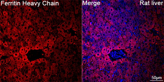
Confocal imaging of paraffin-embedded Rat liver tissue using Ferritin Heavy Chain Rabbit mAb (CAB19544, dilution 1:200) followed by a further incubation with Cy3 Goat Anti-Rabbit IgG (H+L) (AS007, dilution 1:500) (Red). DAPI was used for nuclear staining (Blue). High pressure antigen retrieval performed with 0.01M Citrate Buffer (pH 6.0) prior to IF staining. Objective: 40x.