The FGF1 Antibody (CAB0685) is a high-quality antibody developed for reliable detection and analysis of target proteins. FGF1 is a key player in cell growth, differentiation, and tissue repair, making it a crucial target for studies in developmental biology, cancer research, and regenerative medicine.This polyclonal antibody, produced in rabbits, is highly specific to human FGF1 and has been validated for use in Western blot applications. It effectively detects FGF1 protein, allowing for detailed analysis in different cell types and tissues. Researchers can rely on this antibody to accurately measure FGF1 levels and investigate its function in various biological contexts.
This antibody is validated for use in WB, IHC-P, IF/ICC, ELISA applications and has demonstrated reactivity against Human, Mouse, Rat samples.
Product Name:
FGF1 Antibody
SKU:
CAB0685
Size:
20μL, 100μL
Reactivity:
Human, Mouse, Rat
Conjugate:
Unconjugated
Immunogen:
Recombinant protein (or fragment).This information is considered to be commercially sensitive.
The protein encoded by this gene is a member of the fibroblast growth factor (FGF) family. FGF family members possess broad mitogenic and cell survival activities, and are involved in a variety of biological processes, including embryonic development, cell growth, morphogenesis, tissue repair, tumor growth and invasion. This protein functions as a modifier of endothelial cell migration and proliferation, as well as an angiogenic factor. It acts as a mitogen for a variety of mesoderm- and neuroectoderm-derived cells in vitro, thus is thought to be involved in organogenesis. Multiple alternatively spliced variants encoding different isoforms have been described.
Purification Method
Affinity purification
Gene ID
2246
RRID
AB_2757336
Buffer Information
Store at -20℃. Avoid freeze / thaw cycles. Buffer: PBS containing 50% glycerol, preserved with proclin300 or sodium azide, pH 7.3.
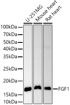
Western blot analysis of various lysates using FGF1 Rabbit pAb (CAB0685) at 1:1000 dilution. Secondary antibody: HRP-conjugated Goat anti-Rabbit IgG (H+L) (CABS014) at 1:10000 dilution. Lysates/proteins: 25μg per lane. Blocking buffer: 3% nonfat dry milk in TBST. Detection: ECL Basic Kit (AbGn00020). Exposure time: 90s.
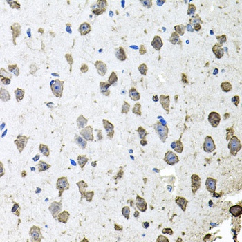
Immunohistochemistry analysis of paraffin-embedded Mouse brain using FGF1 Rabbit pAb (CAB0685) at dilution of 1:100 (40x lens). Microwave antigen retrieval performed with 0.01M PBS Buffer (pH 7.2) prior to IHC staining.
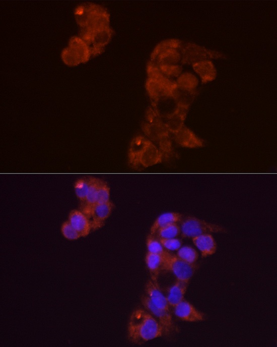
Immunofluorescence analysis of HepG2 cells using FGF1 Rabbit pAb (CAB0685) at dilution of 1:50 (40x lens). Secondary antibody: Cy3-conjugated Goat anti-Rabbit IgG (H+L) (CABS007) at 1:500 dilution. Blue: DAPI for nuclear staining.