The Ambra1 Antibody (CAB1083) is a high-quality antibody developed for reliable detection and analysis of target proteins. Enables GTPase binding activity and ubiquitin protein ligase binding activity. Involved in macroautophagy; positive regulation of phosphatidylinositol 3-kinase activity; and response to mitochondrial depolarisation. Located in cytosol. Colocalizes with mitochondrion. Biomarker of multiple system atrophy.
This antibody is validated for use in WB, IHC-P, IF/ICC, ELISA applications and has demonstrated reactivity against Human, Mouse, Rat samples.
Product Name:
Ambra1 Antibody
SKU:
CAB1083
Size:
100μL, 20μL
Reactivity:
Human, Mouse, Rat
Conjugate:
Unconjugated
Immunogen:
Recombinant protein (or fragment).This information is considered to be commercially sensitive.
Tested Applications:
WBIHC-PIF/ICCELISA
Recommended Dilution:
WB
1:500 - 1:1000
IHC-P
1:50 - 1:200
IF/ICC
1:50 - 1:200
ELISA
Recommended starting concentration is 1 μg/mL. Please optimize the concentration based on your specific assay requirements.
Synonyms:
DCAF3, WDR94, Ambra1
Positive Sample:
PANC-1
Cellular Localization:
Cytoplasmic Vesicle, Autophagosome.
Calculated MW:
143kDa
Observed MW:
130-150kDa
Enables GTPase binding activity and ubiquitin protein ligase binding activity. Involved in macroautophagy; positive regulation of phosphatidylinositol 3-kinase activity; and response to mitochondrial depolarisation. Located in cytosol. Colocalizes with mitochondrion. Biomarker of multiple system atrophy.
Purification Method
Affinity purification
Gene ID
55626
RRID
AB_2758254
Buffer Information
Store at -20℃. Avoid freeze / thaw cycles. Buffer: PBS containing 50% glycerol, preserved with proclin300 or sodium azide, pH 7.3.
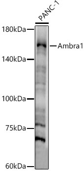
Western blot analysis of lysates from PANC-1 cells, using Ambra1 Rabbit pAb (CAB1083) at 1:800 dilution. Secondary antibody: HRP-conjugated Goat anti-Rabbit IgG (H+L) (AS014) at 1:10000 dilution. Lysates/proteins: 25μg per lane. Blocking buffer: 3% nonfat dry milk in TBST. Detection: ECL Enhanced Kit (AbGn00021). Exposure time: 30s.
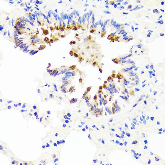
Immunohistochemistry analysis of paraffin-embedded Rat lung using Ambra1 Rabbit pAb (CAB1083) at dilution of 1:100 (40x lens). Microwave antigen retrieval performed with 0.01M PBS Buffer (pH 7.2) prior to IHC staining.
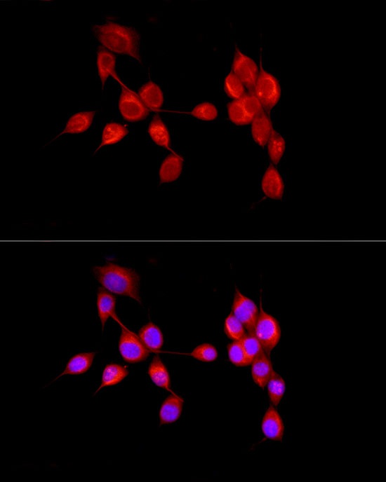
Immunofluorescence analysis of NIH/3T3 cells using Ambra1 Rabbit pAb (CAB1083) at dilution of 1:50 (40x lens). Secondary antibody: Cy3-conjugated Goat anti-Rabbit IgG (H+L) (AS007) at 1:500 dilution. Blue: DAPI for nuclear staining.