The FUNDC1 Antibody (CAB16318) is a high-quality antibody developed for reliable detection and analysis of target proteins. This antibody, generated in rabbits, exhibits high specificity and sensitivity when used in Western blot applications with human samples. By binding to FUNDC1, it enables the precise detection and analysis of this protein in a variety of cell types.FUNDC1, a mitochondrial outer membrane protein, is involved in maintaining mitochondrial homeostasis and quality control through selective autophagy. Its role in mitophagy, the process of degrading damaged mitochondria, makes it a crucial target for research into cell metabolism, aging, and neurodegenerative diseases.
This antibody is validated for use in WB, IHC-P, ELISA applications and has demonstrated reactivity against Human, Mouse samples.
Product Name:
FUNDC1 Antibody
SKU:
CAB16318
Size:
20μL, 100μL
Reactivity:
Human, Mouse
Conjugate:
Unconjugated
Immunogen:
Synthetic peptide. This information is considered to be commercially sensitive.
Recommended starting concentration is 1 μg/mL. Please optimize the concentration based on your specific assay requirements.
Synonyms:
FUNDC1
Positive Sample:
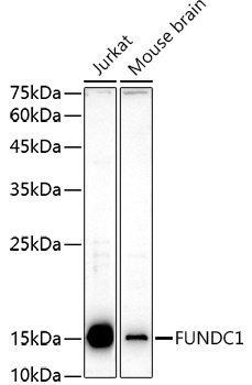
Jurkat, Mouse brain
Cellular Localization:
Mitochondrial Outer Membrane.
Calculated MW:
17kDa
Observed MW:
17kDa
This gene encodes a protein with a FUN14 superfamily domain. The function of the encoded protein is not known.
Purification Method
Affinity purification
Gene ID
139341
RRID
AB_2769533
Buffer Information
Store at -20℃. Avoid freeze / thaw cycles. Buffer: PBS containing 50% glycerol, preserved with proclin300 or sodium azide, pH 7.3.
Western blot analysis of various lysates using FUNDC1 Rabbit pAb (CAB16318) at 1:1000 dilution. Secondary antibody: HRP-conjugated Goat anti-Rabbit IgG (H+L) (CABS014) at 1:10000 dilution. Lysates/proteins: 25μg per lane. Blocking buffer: 3% nonfat dry milk in TBST. Detection: ECL Enhanced Kit (AbGn00021). Exposure time: 180s.
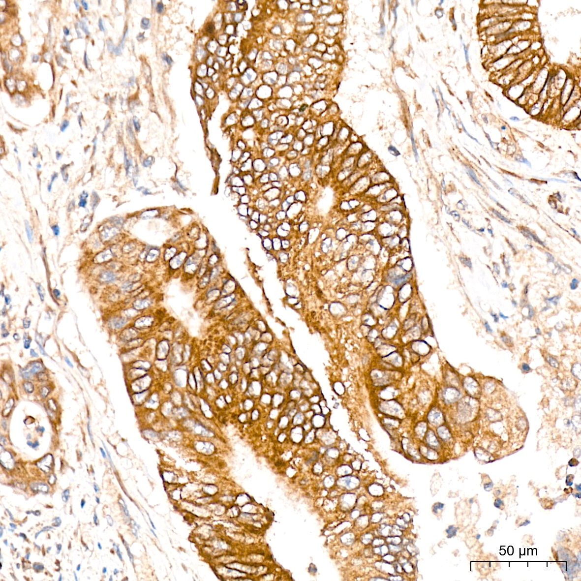
Immunohistochemistry analysis of paraffin-embedded Human colon carcinoma tissue using FUNDC1 Rabbit pAb (CAB16318) at a dilution of 1:100 (40x lens). High pressure antigen retrieval was performed with 0.01 M citrate buffer (pH 6.0) prior to IHC staining.