Vitamin D Binding protein Monoclonal Antibody (CAB21977)
The Vitamin D Binding protein Monoclonal Antibody (CAB21977) is a high-quality antibody developed for reliable detection and analysis of target proteins. This antibody, produced using advanced monoclonal antibody technology, shows high specificity and sensitivity for VDBP in human samples, making it ideal for various research applications.Vitamin D binding protein, also known as GC protein, is involved in the transport of Vitamin D metabolites in the bloodstream, as well as modulating immune responses and inflammation. Dysregulation of VDBP has been implicated in various diseases, including osteoporosis, cardiovascular disease, and certain types of cancer.
This antibody is validated for use in WB, ELISA applications and has demonstrated reactivity against Human, Mouse, Rat samples.
Product Name:
Vitamin D Binding protein Monoclonal Antibody
SKU:
CAB21977
Size:
20μL, 100μL
Reactivity:
Human, Mouse, Rat
Clone Number:
ARC53914
Conjugate:
Unconjugated
Immunogen:
Recombinant protein (or fragment).This information is considered to be commercially sensitive.
Recommended starting concentration is 1 μg/mL. Please optimize the concentration based on your specific assay requirements.
Synonyms:
DBP, VDB, GRD3, VDBG, VDBP, GcMAF, DBP/GC, Gc-MAF, DBP-maf, HEL-S-51, Vitamin D Binding protein
Positive Sample:
Mouse liver, Rat plasma
Cellular Localization:
Secreted.
Calculated MW:
53kDa
Observed MW:
53kDa
The protein encoded by this gene belongs to the albumin gene family. It is a multifunctional protein found in plasma, ascitic fluid, cerebrospinal fluid and on the surface of many cell types. It binds to vitamin D and its plasma metabolites and transports them to target tissues. Alternatively spliced transcript variants encoding different isoforms have been found for this gene.
Purification Method
Affinity purification
Gene ID
2638
Buffer Information
Store at -20℃. Avoid freeze / thaw cycles. Buffer: PBS containing 50% glycerol and 0.05% BSA, preserved with proclin300 or sodium azide, pH 7.3.
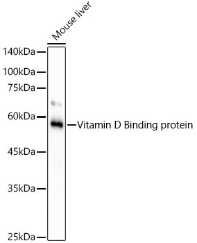
Western blot analysis of lysates from mouse liver, using Vitamin D Binding protein Rabbit mAb (CAB21977) at 1:1000 dilution. Secondary antibody: HRP-conjugated Goat anti-Rabbit IgG (H+L) (CABS014) at 1:10000 dilution. Lysates/proteins: 25μg per lane. Blocking buffer: 3% nonfat dry milk in TBST. Detection: ECL Basic Kit (AbGn00020). Exposure time: 90s.
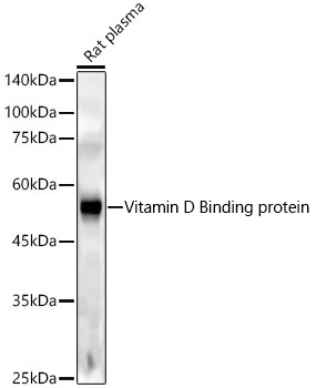
Western blot analysis of lysates from rat plasma, using Vitamin D Binding protein Rabbit mAb (CAB21977) at 1:1000 dilution. Secondary antibody: HRP-conjugated Goat anti-Rabbit IgG (H+L) (CABS014) at 1:10000 dilution. Lysates/proteins: 25μg per lane. Blocking buffer: 3% nonfat dry milk in TBST. Detection: ECL Enhanced Kit (AbGn00021). Exposure time: 180s.
 at 1:1000 dilution. Secondary antibody: HRP Goat Anti-Rabbit IgG (H+L) at 1:10000 dilution. Lysates/proteins: 25μg per lane. Blocking buffer: 3% nonfat dry milk in TBST.")
 at 1:1000 dilution. Secondary antibody: HRP Goat Anti-Rabbit IgG (H+L) at 1:10000 dilution. Lysates/proteins: 25μg per lane. Blocking buffer: 3% nonfat dry milk in TBST.")


 ELISA Kit (MOEB1707)")
 ELISA Kit (RTEB1215)")