The GFAP Antibody (CAB0237) is a high-quality antibody developed for reliable detection and analysis of target proteins. This antibody, produced in rabbits, exhibits high reactivity with human samples and has been rigorously validated for use in techniques such as Western blotting.GFAP is crucial for maintaining the structural integrity and function of astrocytes, which are essential for supporting neuronal health and function in the brain and spinal cord. Abnormal levels or expression patterns of GFAP have been linked to various neurological disorders, including Alzheimer's disease, traumatic brain injury, and multiple sclerosis.
This antibody is validated for use in WB, IHC-P, IF/ICC, ELISA, IF-P applications and has demonstrated reactivity against Human, Mouse, Rat samples.
Product Name:
GFAP Antibody
SKU:
CAB0237
Size:
20μL, 100μL
Reactivity:
Human, Mouse, Rat
Conjugate:
Unconjugated
Immunogen:
Synthetic peptide. This information is considered to be commercially sensitive.
Sequence:
MERR RITS AARR SYVS SGEM MVGG LAPG RRLG PGTR LSLA RMPP PLPT RVDF SLAG ALNA GFKE TRAS ERAE MME
Tested Applications:
WBIHC-PIF/ICCELISAIF-P
Recommended Dilution:
WB
1:500 - 1:1000
IF/ICC
1:50 - 1:200
IF-P
1:50 - 1:200
IHC-P
1:50 - 1:200
ELISA
Recommended starting concentration is 1 μg/mL. Please optimize the concentration based on your specific assay requirements.
Synonyms:
ALXDRD, GFAP
Positive Sample:
Mouse brain
Cellular Localization:
Cytoplasm.
Calculated MW:
50kDa
Observed MW:
50kDa
This gene encodes one of the major intermediate filament proteins of mature astrocytes. It is used as a marker to distinguish astrocytes from other glial cells during development. Mutations in this gene cause Alexander disease, a rare disorder of astrocytes in the central nervous system. Alternative splicing results in multiple transcript variants encoding distinct isoforms.
Purification Method
Affinity purification
Gene ID
2670
RRID
AB_2757050
Buffer Information
Store at -20℃. Avoid freeze / thaw cycles. Buffer: PBS containing 50% glycerol, preserved with proclin300 or sodium azide, pH 7.3.
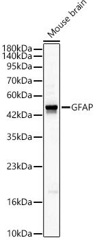
Western blot analysis of lysates from Mouse brain, using GFAP Rabbit pAb (CAB0237) at 1:1000 dilution. Secondary antibody: HRP-conjugated Goat anti-Rabbit IgG (H+L) (CABS014) at 1:10000 dilution. Lysates/proteins: 25μg per lane. Blocking buffer: 3% nonfat dry milk in TBST. Detection: ECL Basic Kit (AbGn00020). Exposure time: 60s.
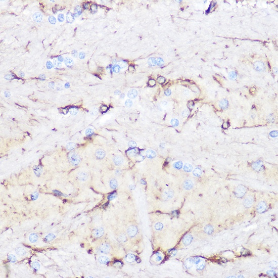
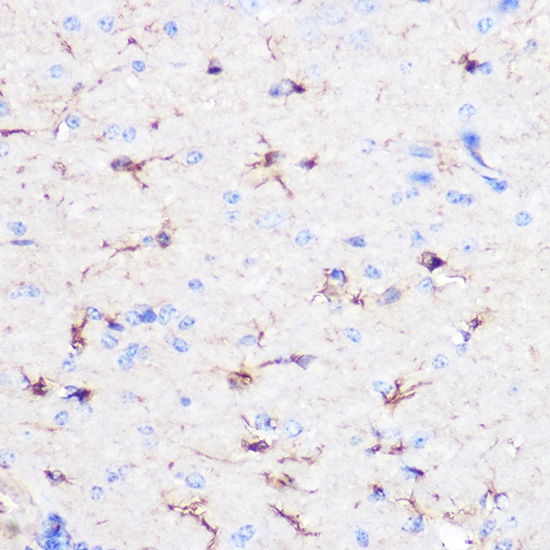
Immunohistochemistry analysis of paraffin-embedded Rat brain using GFAP Rabbit pAb (CAB0237) at dilution of 1:100 (40x lens). Microwave antigen retrieval performed with 0.01M Tris/EDTA Buffer (pH 9.0) prior to IHC staining.
Immunohistochemistry analysis of paraffin-embedded Mouse brain using GFAP Rabbit pAb (CAB0237) at dilution of 1:100 (40x lens). Microwave antigen retrieval performed with 0.01M Tris/EDTA Buffer (pH 9.0) prior to IHC staining.
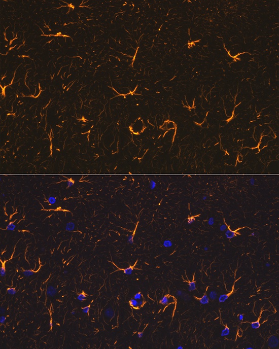
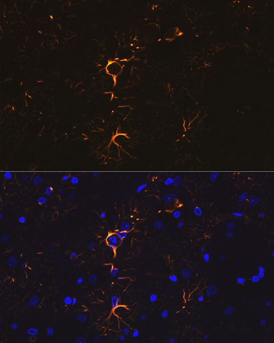
Immunofluorescence analysis of paraffin-embedded rat brain using GFAP Rabbit pAb (CAB0237) at dilution of 1:100 (40x lens). Secondary antibody: Cy3-conjugated Goat anti-Rabbit IgG (H+L) (CABS007) at 1:500 dilution. Blue: DAPI for nuclear staining.
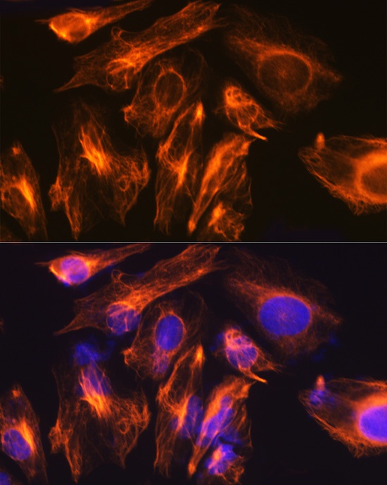
Immunofluorescence analysis of U-251MG cells using GFAP Rabbit pAb (CAB0237) at dilution of 1:100 (40x lens). Secondary antibody: Cy3-conjugated Goat anti-Rabbit IgG (H+L) (CABS007) at 1:500 dilution. Blue: DAPI for nuclear staining.
Immunofluorescence analysis of paraffin-embedded rat brain using GFAP Rabbit pAb (CAB0237) at dilution of 1:100 (40x lens). Secondary antibody: Cy3-conjugated Goat anti-Rabbit IgG (H+L) (CABS007) at 1:500 dilution. Blue: DAPI for nuclear staining.