The GLDC Antibody (CAB9933) is a high-quality antibody developed for reliable detection and analysis of target proteins. This antibody, produced in rabbits, exhibits high specificity and sensitivity for detecting GLDC in human samples, making it an excellent choice for Western blot applications.GLDC plays a vital role in the breakdown of glycine, an essential amino acid involved in numerous cellular processes. Dysregulation of GLDC has been associated with a variety of metabolic disorders and neurological conditions, making it a key target for research in these areas.
This antibody is validated for use in WB, IF/ICC, IP, ELISA applications and has demonstrated reactivity against Human, Mouse, Rat samples.
Product Name:
GLDC Antibody
SKU:
CAB9933
Size:
20μL, 100μL
Reactivity:
Human, Mouse, Rat
Conjugate:
Unconjugated
Immunogen:
Recombinant protein (or fragment).This information is considered to be commercially sensitive.
0.5μg-4μg antibody for 200μg-400μg extracts of whole cells
ELISA
Recommended starting concentration is 1 μg/mL. Please optimize the concentration based on your specific assay requirements.
Synonyms:
GCE, GCSP, HYGN1, GLDC
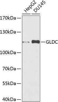
Positive Sample:
HepG2, DU145
Cellular Localization:
Mitochondrion.
Calculated MW:
113kDa
Observed MW:
113kDa
Degradation of glycine is brought about by the glycine cleavage system, which is composed of four mitochondrial protein components: P protein (a pyridoxal phosphate-dependent glycine decarboxylase), H protein (a lipoic acid-containing protein), T protein (a tetrahydrofolate-requiring enzyme), and L protein (a lipoamide dehydrogenase). The protein encoded by this gene is the P protein, which binds to glycine and enables the methylamine group from glycine to be transferred to the T protein. Defects in this gene are a cause of nonketotic hyperglycinemia (NKH).
Purification Method
Affinity purification
Gene ID
2731
RRID
AB_2769610
Buffer Information
Store at -20℃. Avoid freeze / thaw cycles. Buffer: PBS containing 50% glycerol, preserved with proclin300 or sodium azide, pH 7.3.
Western blot analysis of various lysates using GLDC Rabbit pAb (CAB9933) at 1:1000 dilution. Secondary antibody: HRP-conjugated Goat anti-Rabbit IgG (H+L) (CABS014) at 1:10000 dilution. Lysates/proteins: 25μg per lane. Blocking buffer: 3% nonfat dry milk in TBST. Detection: ECL Basic Kit (AbGn00020). Exposure time: 90s.
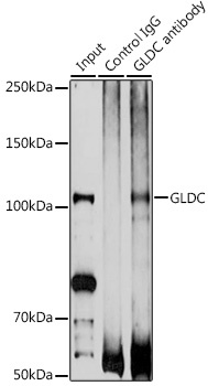
Immunoprecipitation analysis of 200 μg extracts of DU145 cells using 3 μg GLDC antibody (CAB9933). Western blot was performed from the immunoprecipitate using GLDC antibody (CAB9933) at a dilution of 1:1000.


")


