The Glycophorin C (GYPC) Antibody (CAB1232) is a high-quality antibody developed for reliable detection and analysis of target proteins. This antibody, produced in rabbits, demonstrates high specificity and sensitivity for detecting Glycophorin-C in human samples, making it an essential reagent for Western blot analysis.Glycophorin-C, a glycoprotein found on the surface of red blood cells, plays a crucial role in maintaining cell shape and stability, as well as mediating cell-cell interactions. Dysregulation of Glycophorin-C expression has been linked to various blood disorders and conditions, making it a target of interest for researchers in hematology and related fields.
This antibody is validated for use in WB, ELISA applications and has demonstrated reactivity against Human, Mouse samples.
Product Name:
Glycophorin C (GYPC) Antibody
SKU:
CAB1232
Size:
20μL, 100μL
Reactivity:
Human, Mouse
Conjugate:
Unconjugated
Immunogen:
Recombinant protein (or fragment).This information is considered to be commercially sensitive.
Cell Membrane, Single-Pass Type Iii Membrane Protein.
Calculated MW:
14kDa
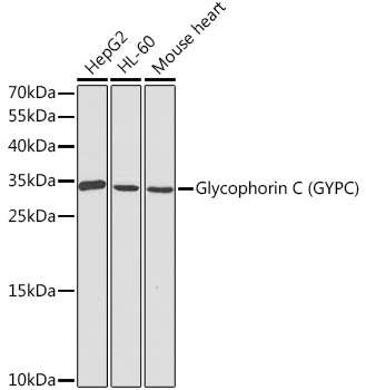
Observed MW:
34kDa
Glycophorin C (GYPC) is an integral membrane glycoprotein. It is a minor species carried by human erythrocytes, but plays an important role in regulating the mechanical stability of red cells. A number of glycophorin C mutations have been described. The Gerbich and Yus phenotypes are due to deletion of exon 3 and 2, respectively. The Webb and Duch antigens, also known as glycophorin D, result from single point mutations of the glycophorin C gene. The glycophorin C protein has very little homology with glycophorins A and B. Alternate splicing results in multiple transcript variants.
Purification Method
Affinity purification
Gene ID
2995
RRID
AB_2759172
Buffer Information
Store at -20℃. Avoid freeze / thaw cycles. Buffer: PBS containing 50% glycerol, preserved with proclin300 or sodium azide, pH 7.3.
Western blot analysis of various lysates using Glycophorin C (GYPC) Rabbit pAb (CAB1232) at 1:1000 dilution. Secondary antibody: HRP-conjugated Goat anti-Rabbit IgG (H+L) (CABS014) at 1:10000 dilution. Lysates/proteins: 25μg per lane. Blocking buffer: 3% nonfat dry milk in TBST. Detection: ECL Basic Kit (AbGn00020). Exposure time: 90s.