The GNL3 Antibody (CAB6459) is a high-quality antibody developed for reliable detection and analysis of target proteins. This antibody, raised in rabbits, offers high specificity and sensitivity for detecting GNL3 in human samples, making it ideal for use in Western blot applications.GNL3 is a nucleolar protein that has been implicated in various biological processes, including ribosome biogenesis, cell cycle regulation, and cancer development. Its overexpression has been observed in many types of cancer, suggesting a potential role in tumorigenesis.
This antibody is validated for use in WB, IF/ICC, IP, ELISA applications and has demonstrated reactivity against Human samples.
Product Name:
GNL3 Antibody
SKU:
CAB6459
Size:
20μL, 100μL
Reactivity:
Human
Conjugate:
Unconjugated
Immunogen:
Recombinant protein (or fragment).This information is considered to be commercially sensitive.
0.5μg-4μg antibody for 200μg-400μg extracts of whole cells
ELISA
Recommended starting concentration is 1 μg/mL. Please optimize the concentration based on your specific assay requirements.
Synonyms:
NS, E2IG3, NNP47, C77032, GNL3
Positive Sample:
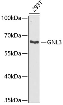
293T
Cellular Localization:
Nucleus, Nucleolus.
Calculated MW:
62kDa
Observed MW:
62kDa
The protein encoded by this gene may interact with p53 and may be involved in tumorigenesis. The encoded protein also appears to be important for stem cell proliferation. This protein is found in both the nucleus and nucleolus. Three transcript variants encoding two different isoforms have been found for this gene.
Purification Method
Affinity purification
Gene ID
26354
RRID
AB_2767061
Buffer Information
Store at -20℃. Avoid freeze / thaw cycles. Buffer: PBS containing 50% glycerol, preserved with proclin300 or sodium azide, pH 7.3.
Western blot analysis of lysates from 293T cells, using GNL3 Rabbit pAb (CAB6459) at 1:1000 dilution. Secondary antibody: HRP-conjugated Goat anti-Rabbit IgG (H+L) (CABS014) at 1:10000 dilution. Lysates/proteins: 25μg per lane. Blocking buffer: 3% nonfat dry milk in TBST. Detection: ECL Basic Kit (AbGn00020). Exposure time: 90s.
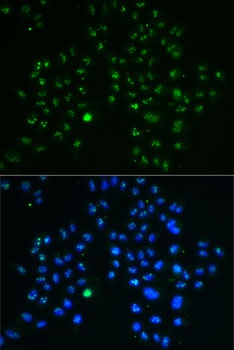
Immunofluorescence analysis of MCF7 cells using GNL3 Rabbit pAb (CAB6459). Secondary antibody: Cy3-conjugated Goat anti-Rabbit IgG (H+L) (CABS007) at 1:500 dilution. Blue: DAPI for nuclear staining.