The GRASP65 Monoclonal Antibody (CAB2316) is a high-quality antibody developed for reliable detection and analysis of target proteins. This polyclonal antibody, generated in rabbits, exhibits strong reactivity with human samples and has been validated for use in Western blot applications.GRASP65 plays a critical role in maintaining the structural integrity of the Golgi apparatus and facilitating the proper sorting and trafficking of proteins to their respective destinations within the cell.
This antibody is validated for use in WB, IF/ICC, ELISA applications and has demonstrated reactivity against Human, Mouse, Rat samples.
Product Name:
GRASP65 Monoclonal Antibody
SKU:
CAB2316
Size:
20μL, 100μL
Reactivity:
Human, Mouse, Rat
Clone Number:
ARC1911
Conjugate:
Unconjugated
Immunogen:
Synthetic peptide. This information is considered to be commercially sensitive.
Sequence:
Email for sequence
Tested Applications:
WBIF/ICCELISA
Recommended Dilution:
WB
1:500 - 1:1000
IF/ICC
1:50 - 1:200
ELISA
Recommended starting concentration is 1 μg/mL. Please optimize the concentration based on your specific assay requirements.
The Golgi complex plays a key role in the sorting and modification of proteins exported from the endoplasmic reticulum. The protein encoded by this gene is a membrane protein involved in establishing the stacked structure of the Golgi apparatus. It is a caspase-3 substrate, and cleavage of this encoded protein contributes to Golgi fragmentation in apoptosis. This encoded protein can form a complex with the Golgi matrix protein GOLGA2, and this complex binds to the vesicle docking protein p115. Alternative splicing results in multiple transcript variants of this gene.
Purification Method
Affinity purification
Gene ID
64689
RRID
AB_2862993
Buffer Information
Store at -20℃. Avoid freeze / thaw cycles. Buffer: PBS containing 50% glycerol and 0.05% BSA, preserved with proclin300 or sodium azide, pH 7.3.
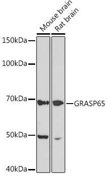
Western blot analysis of various lysates using GRASP65 Rabbit mAb (CAB2316) at 1:1000 dilution. Secondary antibody: HRP-conjugated Goat anti-Rabbit IgG (H+L) (CABS014) at 1:10000 dilution. Lysates/proteins: 25μg per lane. Blocking buffer: 3% nonfat dry milk in TBST. Detection: ECL Basic Kit (AbGn00020). Exposure time: 10s.
Western blot analysis of various lysates using GRASP65 Rabbit mAb (CAB2316) at 1:1000 dilution. Secondary antibody: HRP-conjugated Goat anti-Rabbit IgG (H+L) (CABS014) at 1:10000 dilution. Lysates/proteins: 25μg per lane. Blocking buffer: 3% nonfat dry milk in TBST. Detection: ECL Basic Kit (AbGn00020). Exposure time: 3min.
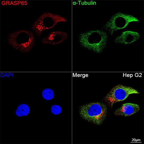
Confocal imaging of Hep G2 cells using GRASP65 Rabbit mAb (CAB2316, dilution 1:100) followed by a further incubation with Cy3 Goat Anti-Rabbit IgG (H+L) (CABS007, dilution 1:500) (Red). The cells were counterstained with α-Tubulin Mouse mAb (AC012, dilution 1:400) followed by incubation with ABflo® 488-conjugated Goat Anti-Mouse IgG (H+L) Ab (CABS076, dilution 1:500) (Green). DAPI was used for nuclear staining (Blue). Objective: 100x.


 ELISA Kit (HUFI03087)")