The GPS1 Antibody (CAB6917) is a high-quality antibody developed for reliable detection and analysis of target proteins. This antibody, produced using rabbit immunization, is highly reactive with human samples and has been validated for use in Western blot applications. It binds to the GPS1 protein, allowing for accurate detection and analysis in a variety of cell types, making it an essential tool for studies in molecular biology and gene regulation.GPS1, also known as G Protein Pathway Suppressor 1, is a key player in the regulation of gene expression by inhibiting the G protein signaling pathway.
This antibody is validated for use in WB, IHC-P, IF/ICC, ELISA applications and has demonstrated reactivity against Human, Mouse, Rat samples.
Product Name:
GPS1 Antibody
SKU:
CAB6917
Size:
20μL, 100μL
Reactivity:
Human, Mouse, Rat
Conjugate:
Unconjugated
Immunogen:
Recombinant protein (or fragment).This information is considered to be commercially sensitive.
Recommended starting concentration is 1 μg/mL. Please optimize the concentration based on your specific assay requirements.
Synonyms:
CSN1, SGN1, COPS1, GPS1
Positive Sample:
293T, SKOV3, Mouse brain
Cellular Localization:
Cytoplasm, Nucleus.
Calculated MW:
56kDa
Observed MW:
55kDa
This gene is known to suppress G-protein and mitogen-activated signal transduction in mammalian cells. The encoded protein shares significant similarity with Arabidopsis FUS6, which is a regulator of light-mediated signal transduction in plant cells.
Purification Method
Affinity purification
Gene ID
2873
RRID
AB_2767476
Buffer Information
Store at -20℃. Avoid freeze / thaw cycles. Buffer: PBS containing 50% glycerol, preserved with proclin300 or sodium azide, pH 7.3.
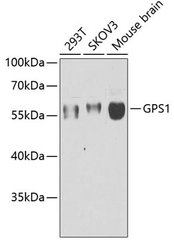
Western blot analysis of various lysates using GPS1 Rabbit pAb (CAB6917) at 1:1000 dilution. Secondary antibody: HRP-conjugated Goat anti-Rabbit IgG (H+L) (CABS014) at 1:10000 dilution. Lysates/proteins: 25μg per lane. Blocking buffer: 3% nonfat dry milk in TBST. Detection: ECL Enhanced Kit (AbGn00021). Exposure time: 90s.
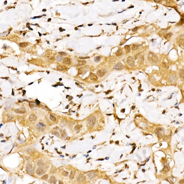
Immunohistochemistry analysis of paraffin-embedded Human breast cancer using GPS1 Rabbit pAb (CAB6917) at dilution of 1:200 (40x lens). High pressure antigen retrieval performed with 0.01M Citrate buffer (pH 6.0) prior to IHC staining.
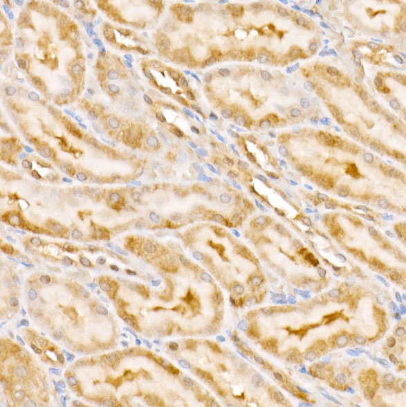
Immunohistochemistry analysis of paraffin-embedded Rat kidney using GPS1 Rabbit pAb (CAB6917) at dilution of 1:200 (40x lens). High pressure antigen retrieval performed with 0.01M Citrate buffer (pH 6.0) prior to IHC staining.
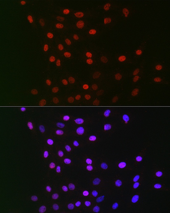
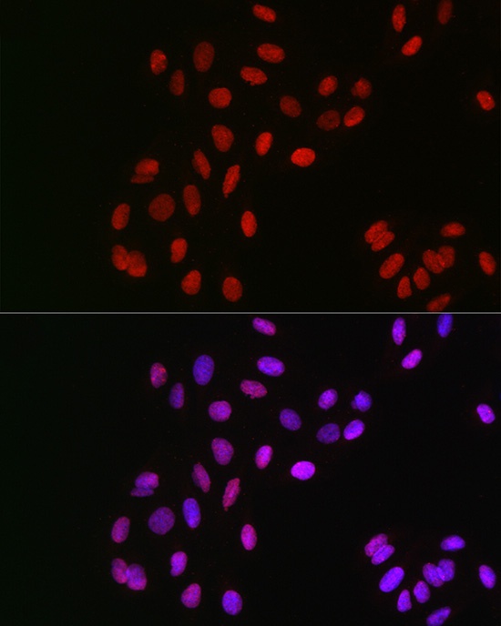
Immunofluorescence analysis of C6 cells using GPS1 Rabbit pAb (CAB6917) at dilution of 1:100 (40x lens). Secondary antibody: Cy3-conjugated Goat anti-Rabbit IgG (H+L) (CABS007) at 1:500 dilution. Blue: DAPI for nuclear staining.
Immunofluorescence analysis of U2OS cells using GPS1 Rabbit pAb (CAB6917) at dilution of 1:100 (40x lens). Secondary antibody: Cy3-conjugated Goat anti-Rabbit IgG (H+L) (CABS007) at 1:500 dilution. Blue: DAPI for nuclear staining.