The GRK1 Antibody (CAB2966) is a high-quality antibody developed for reliable detection and analysis of target proteins. Rhodopsin kinase plays a key role in the phosphorylation of rhodopsin, a light-sensitive receptor protein in the retina, which is essential for the adaptation of the eye to varying light conditions.Raised in rabbits, this antibody is highly specific and reactive with human samples, making it an ideal choice for immunohistochemistry and Western blot applications. By binding to rhodopsin kinase, this antibody enables the detection and analysis of this crucial enzyme in various cell types, providing valuable insights into the mechanisms underlying vision and potential therapeutic targets for retinal diseases.
This antibody is validated for use in WB, IHC-P, ELISA applications and has demonstrated reactivity against Human, Mouse, Rat samples.
Product Name:
GRK1 Antibody
SKU:
CAB2966
Size:
20μL, 100μL
Reactivity:
Human, Mouse, Rat
Conjugate:
Unconjugated
Immunogen:
Recombinant protein (or fragment).This information is considered to be commercially sensitive.
Recommended starting concentration is 1 μg/mL. Please optimize the concentration based on your specific assay requirements.
Synonyms:
RK, RHOK, GPRK1, GRK1
Positive Sample:
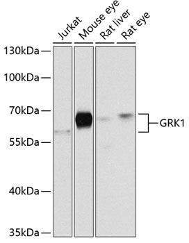
Jurkat, Mouse eye, Rat liver, Rat eye
Cellular Localization:
Lipid-Anchor, Membrane.
Calculated MW:
64kDa
Observed MW:
64kDa
This gene encodes a member of the guanine nucleotide-binding protein (G protein)-coupled receptor kinase subfamily of the Ser/Thr protein kinase family. The protein phosphorylates rhodopsin and initiates its deactivation. Defects in GRK1 are known to cause Oguchi disease 2 (also known as stationary night blindness Oguchi type-2).
Purification Method
Affinity purification
Gene ID
6011
RRID
AB_2764779
Buffer Information
Store at -20℃. Avoid freeze / thaw cycles. Buffer: PBS containing 50% glycerol, preserved with proclin300 or sodium azide, pH 7.3.
Western blot analysis of various lysates using GRK1 Rabbit pAb (CAB2966) at 1:1000 dilution. Secondary antibody: HRP-conjugated Goat anti-Rabbit IgG (H+L) (CABS014) at 1:10000 dilution. Lysates/proteins: 25μg per lane. Blocking buffer: 3% nonfat dry milk in TBST. Detection: ECL Basic Kit (AbGn00020). Exposure time: 30s.
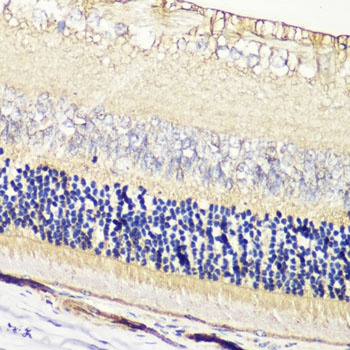
Immunohistochemistry analysis of paraffin-embedded Rat retina using GRK1 Rabbit pAb (CAB2966) at dilution of 1:200 (40x lens). Microwave antigen retrieval performed with 0.01M PBS Buffer (pH 7.2) prior to IHC staining.