The GSDMD Antibody (CAB18281) is a high-quality antibody developed for reliable detection and analysis of target proteins. This rabbit-derived antibody is highly specific and reactive with human samples, making it an excellent choice for studies involving GSDMD in Western blot applications.Gasdermin D has emerged as a key player in the inflammatory response, particularly in the context of infectious diseases and immune dysregulation. By targeting GSDMD, researchers can gain insights into the mechanisms underlying pyroptosis and its role in various pathological conditions, including sepsis, inflammatory bowel disease, and cancer.
This antibody is validated for use in WB, IHC-P, IF/ICC, IP, ELISA applications and has demonstrated reactivity against Human, Mouse, Rat samples.
Product Name:
GSDMD Antibody
SKU:
CAB18281
Size:
20μL, 100μL
Reactivity:
Human, Mouse, Rat
Immunogen:
Recombinant protein (or fragment).This information is considered to be commercially sensitive.
Sequence:
Email for sequence
Tested Applications:
WBIHC-PIF/ICCIPELISA
Recommended Dilution:
WB
1:1000 - 1:2000
IP
0.5μg-4μg antibody for 200μg-400μg extracts of whole cells
IF/ICC
1:50 - 1:200
IHC-P
1:50 - 1:200
ELISA
Recommended starting concentration is 1 μg/mL. Please optimize the concentration based on your specific assay requirements.
Synonyms:
DF5L, DFNA5L, FKSG10, GSDMDC1, GSDMD
Positive Sample:
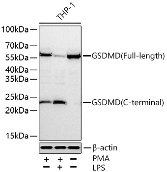
THP-1 treated with PMA and LPS, THP-1 treated with PMA, THP-1
Gasdermin D is a member of the gasdermin family. Members of this family appear to play a role in regulation of epithelial proliferation. Gasdermin D has been suggested to act as a tumor suppressor. Alternatively spliced transcript variants have been described.
Purification Method
Affinity purification
Gene ID
79792
RRID
AB_2862055
Buffer Information
Store at -20℃. Avoid freeze / thaw cycles. Buffer: PBS containing 50% glycerol, preserved with proclin300 or sodium azide, pH 7.3.
Western blot analysis of various lysates using GSDMD Rabbit pAb (CAB18281) at 1:1000 dilution incubated overnight at 4℃. THP-1 cells were treated with PMA(80 nM)at 37 ℃ for 24 hours, LPS(5 μg/mL) at 37 ℃ for 6 hours. Secondary antibody: HRP-conjugated Goat anti-Rabbit IgG (H+L) (CABS014) at 1:10000 dilution. Lysates/proteins: 30 μg per lane. Blocking buffer: 3% nonfat dry milk in TBST. Detection: ECL Basic Kit (AbGn00020). Exposure time: 90 s.


. Blue: DAPI for nuclear staining.")
. Blue: DAPI for nuclear staining.")
 Monoclonal Antibody (CAB23755)")


 ELISA Kit (AEFI00984)")
 ELISA Kit (AEFI00984)")