The GSTT1 Antibody (CAB1287) is a high-quality antibody developed for reliable detection and analysis of target proteins. This antibody, produced in rabbits, is highly specific to human samples and has been validated for use in Western blot applications.GSTT1 is a member of the glutathione S-transferase family, which plays a crucial role in protecting cells from oxidative stress and toxic compounds by catalyzing the conjugation of glutathione to a wide variety of substrates. Dysregulation of GSTT1 activity has been implicated in various diseases, including cancer, neurodegenerative disorders, and cardiovascular diseases.
This antibody is validated for use in WB, IHC-P, IF/ICC, ELISA applications and has demonstrated reactivity against Human, Mouse, Rat samples.
Product Name:
GSTT1 Antibody
SKU:
CAB1287
Size:
20μL, 100μL
Reactivity:
Human, Mouse, Rat
Conjugate:
Unconjugated
Immunogen:
Recombinant protein (or fragment).This information is considered to be commercially sensitive.
Recommended starting concentration is 1 μg/mL. Please optimize the concentration based on your specific assay requirements.
Synonyms:
GSTT1
Positive Sample:
HepG2
Cellular Localization:
Cytoplasm.
Calculated MW:
27kDa
Observed MW:
25kDa
The protein encoded by this gene, glutathione S-transferase (GST) theta 1 (GSTT1), is a member of a superfamily of proteins that catalyze the conjugation of reduced glutathione to a variety of electrophilic and hydrophobic compounds. Human GSTs can be divided into five main classes: alpha, mu, pi, theta, and zeta. The theta class includes GSTT1, GSTT2, and GSTT2B. GSTT1 and GSTT2/GSTT2B share 55% amino acid sequence identity and may play a role in human carcinogenesis. The GSTT1 gene is haplotype-specific and is absent from 38% of the population. Alternative splicing of this gene results in multiple transcript variants.
Purification Method
Affinity purification
Gene ID
2952
RRID
AB_2759711
Buffer Information
Store at -20℃. Avoid freeze / thaw cycles. Buffer: PBS containing 50% glycerol, preserved with proclin300 or sodium azide, pH 7.3.
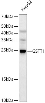
Western blot analysis of lysates from HepG2 cells, using GSTT1 Rabbit pAb (CAB1287) at 1:1000 dilution. Secondary antibody: HRP-conjugated Goat anti-Rabbit IgG (H+L) (CABS014) at 1:10000 dilution. Lysates/proteins: 25μg per lane. Blocking buffer: 3% nonfat dry milk in TBST. Detection: ECL Basic Kit (AbGn00020). Exposure time: 30s.
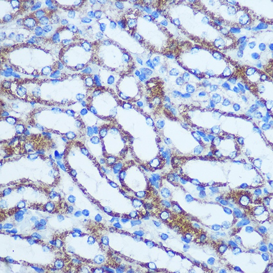
Immunohistochemistry analysis of paraffin-embedded Mouse kidney using GSTT1 Rabbit pAb (CAB1287) at dilution of 1:100 (40x lens). Microwave antigen retrieval performed with 0.01M PBS Buffer (pH 7.2) prior to IHC staining.
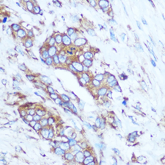
Immunohistochemistry analysis of paraffin-embedded Human breast cancer using GSTT1 Rabbit pAb (CAB1287) at dilution of 1:100 (40x lens). Microwave antigen retrieval performed with 0.01M PBS Buffer (pH 7.2) prior to IHC staining.
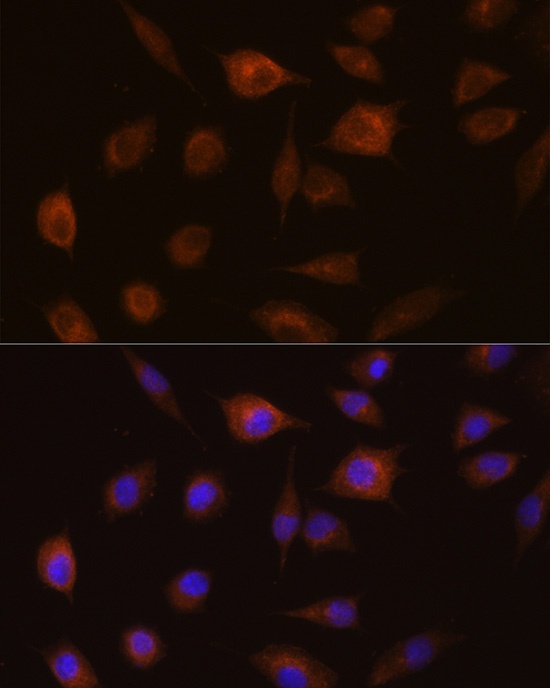
Immunofluorescence analysis of L929 cells using GSTT1 Rabbit pAb (CAB1287) at dilution of 1:100 (40x lens). Secondary antibody: Cy3-conjugated Goat anti-Rabbit IgG (H+L) (CABS007) at 1:500 dilution. Blue: DAPI for nuclear staining.