The HAUS8 Antibody (CAB7847) is a high-quality antibody developed for reliable detection and analysis of target proteins. This antibody, generated in rabbits, exhibits high reactivity with human samples and has been validated for use in Western blot applications. By binding to the HAUS8 protein, this antibody enables the detection and analysis of HAUS8 in various cell types, making it ideal for studies in cell biology, microtubule dynamics, and mitotic spindle formation.The HAUS8 protein is essential for proper mitotic spindle assembly and accurate chromosome segregation during cell division. Dysregulation of HAUS8 function has been linked to defects in cell division and chromosomal instability, which are common features of cancer cells.
This antibody is validated for use in WB, IF/ICC, ELISA applications and has demonstrated reactivity against Human, Mouse samples.
Product Name:
HAUS8 Antibody
SKU:
CAB7847
Size:
20μL, 100μL
Reactivity:
Human, Mouse
Conjugate:
Unconjugated
Immunogen:
Recombinant protein (or fragment).This information is considered to be commercially sensitive.
Recommended starting concentration is 1 μg/mL. Please optimize the concentration based on your specific assay requirements.
Synonyms:
DGT4, HICE1, NY-SAR-48, HAUS8
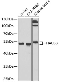
Positive Sample:
Jurkat, NCI-H460, Mouse testis
Cellular Localization:
Cytoplasm, Centrosome, Cytoskeleton, Microtubule Organizing Center, Spindle, Spindle Pole.
Calculated MW:
45kDa
Observed MW:
41kDa
HAUS8 is 1 of 8 subunits of the 390-kD human augmin complex, or HAUS complex. The augmin complex was first identified in Drosophila, and its name comes from the Latin verb 'augmentare,' meaning 'to increase.' The augmin complex is a microtubule-binding complex involved in microtubule generation within the mitotic spindle and is vital to mitotic spindle assembly (Goshima et al., 2008 [PubMed 18443220]; Uehara et al., 2009 [PubMed 19369198]).
Purification Method
Affinity purification
Gene ID
93323
RRID
AB_2769738
Buffer Information
Store at -20℃. Avoid freeze / thaw cycles. Buffer: PBS containing 50% glycerol, preserved with proclin300 or sodium azide, pH 7.3.
Western blot analysis of various lysates using HAUS8 Rabbit pAb (CAB7847) at 1:1000 dilution. Secondary antibody: HRP-conjugated Goat anti-Rabbit IgG (H+L) (CABS014) at 1:10000 dilution. Lysates/proteins: 25μg per lane. Blocking buffer: 3% nonfat dry milk in TBST. Detection: ECL Basic Kit (AbGn00020). Exposure time: 90s.
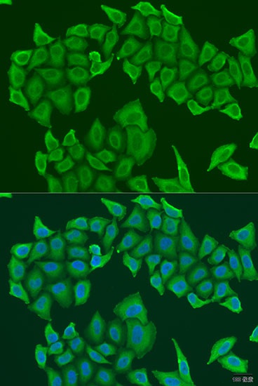
Immunofluorescence analysis of U2OS cells using HAUS8 Rabbit pAb (CAB7847) at dilution of 1:100. Secondary antibody: Cy3-conjugated Goat anti-Rabbit IgG (H+L) (CABS007) at 1:500 dilution. Blue: DAPI for nuclear staining.