The HDAC6 Monoclonal Antibody (CAB22169) is a high-quality antibody developed for reliable detection and analysis of target proteins. This antibody, developed through rigorous research and validation processes, specifically targets and binds to the HDAC6 protein, allowing for precise detection and analysis in various cell types.HDAC6, a member of the histone deacetylase family, plays a key role in many cellular processes, including cell motility, protein degradation, and stress response. Dysregulation of HDAC6 has been linked to various diseases, making it a promising target for therapeutic interventions.
This antibody is validated for use in WB, ELISA applications and has demonstrated reactivity against Human samples.
Product Name:
HDAC6 Monoclonal Antibody
SKU:
CAB22169
Size:
20μL, 100μL
Reactivity:
Human
Clone Number:
ARC55934
Conjugate:
Unconjugated
Immunogen:
Synthetic peptide. This information is considered to be commercially sensitive.
Recommended starting concentration is 1 μg/mL. Please optimize the concentration based on your specific assay requirements.
Synonyms:
HD6, JM21, CPBHM, PPP1R90, HDAC6
Positive Sample:
K-562
Cellular Localization:
Axon Cytoplasm, Caveola, Centrosome, Ciliary Basal Body, Cytoplasm, Cytosol, Histone Deacetylase Complex, Multivesicular Body, Nucleoplasm, Nucleus, Perinuclear Region Of Cytoplasm.
Calculated MW:
131kDa
Observed MW:
160kDa
Histones play a critical role in transcriptional regulation, cell cycle progression, and developmental events. Histone acetylation/deacetylation alters chromosome structure and affects transcription factor access to DNA. The protein encoded by this gene belongs to class II of the histone deacetylase/acuc/apha family. It contains an internal duplication of two catalytic domains which appear to function independently of each other. This protein possesses histone deacetylase activity and represses transcription.
Purification Method
Affinity purification
Gene ID
10013
Buffer Information
Store at -20℃. Avoid freeze / thaw cycles. Buffer: PBS containing 50% glycerol and 0.05% BSA, preserved with proclin300 or sodium azide, pH 7.3.
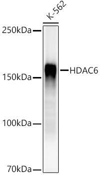
Western blot analysis of lysates from K-562 cells, using HDAC6 Rabbit mAb (CAB22169) at1:2000 dilution. Secondary antibody: HRP-conjugated Goat anti-Rabbit IgG (H+L) (CABS014) at 1:10000 dilution. Lysates/proteins: 25μg per lane. Blocking buffer: 3% nonfat dry milk in TBST. Detection: ECL Enhanced Kit (AbGn00021). Exposure time: 30s.
 at1:2000 dilution. Secondary antibody: HRP Goat Anti-Rabbit IgG (H+L) at 1:10000 dilution. Lysates/proteins: 25μg per lane. Blocking buffer: 3% nonfat dry milk in TBST.")
 at1:2000 dilution. Secondary antibody: HRP Goat Anti-Rabbit IgG (H+L) at 1:10000 dilution. Lysates/proteins: 25μg per lane. Blocking buffer: 3% nonfat dry milk in TBST.")


