The HLA-DRA Monoclonal Antibody (CAB10863) is a high-quality antibody developed for reliable detection and analysis of target proteins. This high-quality antibody, produced in rabbits, is highly specific and reactive with human samples, making it an essential tool for studying immune responses and antigen presentation. Validated for use in applications such as Western blotting, this antibody binds specifically to the HLA-DRA protein, allowing for precise detection and analysis in a variety of cell types.
This antibody is validated for use in WB, IHC-P, ELISA applications and has demonstrated reactivity against Human samples.
Product Name:
HLA-DRA Monoclonal Antibody
SKU:
CAB10863
Size:
20μL, 100μL
Reactivity:
Human
Clone Number:
ARC0518
Conjugate:
Unconjugated
Immunogen:
Synthetic peptide. This information is considered to be commercially sensitive.
Recommended starting concentration is 1 μg/mL. Please optimize the concentration based on your specific assay requirements.
Synonyms:
HLA-DRA1, HLA-DRA
Positive Sample:
Raji
Cellular Localization:
Cell Membrane, Endoplasmic Reticulum Membrane, Endosome Membrane, Golgi Apparatus, Late Endosome Membrane, Lysosome Membrane, Single-Pass Type I Membrane Protein, Trans-Golgi Network Membrane.
Calculated MW:
29kDa
Observed MW:
33kDa
HLA-DRA is one of the HLA class II alpha chain paralogues. This class II molecule is a heterodimer consisting of an alpha and a beta chain, both anchored in the membrane. This molecule is expressed on the surface of various antigen presenting cells such as B lymphocytes, dendritic cells, and monocytes/macrophages, and plays a central role in the immune system and response by presenting peptides derived from extracellular proteins, in particular, pathogen-derived peptides to T cells. The alpha chain is approximately 33-35 kDa and its gene contains 5 exons. Exon 1 encodes the leader peptide, exons 2 and 3 encode the two extracellular domains, and exon 4 encodes the transmembrane domain and the cytoplasmic tail. DRA does not have polymorphisms in the peptide binding part and acts as the sole alpha chain for DRB1, DRB3, DRB4 and DRB5.
Purification Method
Affinity purification
Gene ID
3122
RRID
AB_2861496
Buffer Information
Store at -20℃. Avoid freeze / thaw cycles. Buffer: PBS containing 50% glycerol and 0.05% BSA, preserved with proclin300 or sodium azide, pH 7.3.
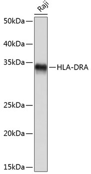
Western blot analysis of lysates from Raji cells, using HLA-DRA Rabbit mAb (CAB10863) at 1:1000 dilution. Secondary antibody: HRP-conjugated Goat anti-Rabbit IgG (H+L) (CABS014) at 1:10000 dilution. Lysates/proteins: 25μg per lane. Blocking buffer: 3% nonfat dry milk in TBST. Detection: ECL Basic Kit (AbGn00020). Exposure time: 1s.
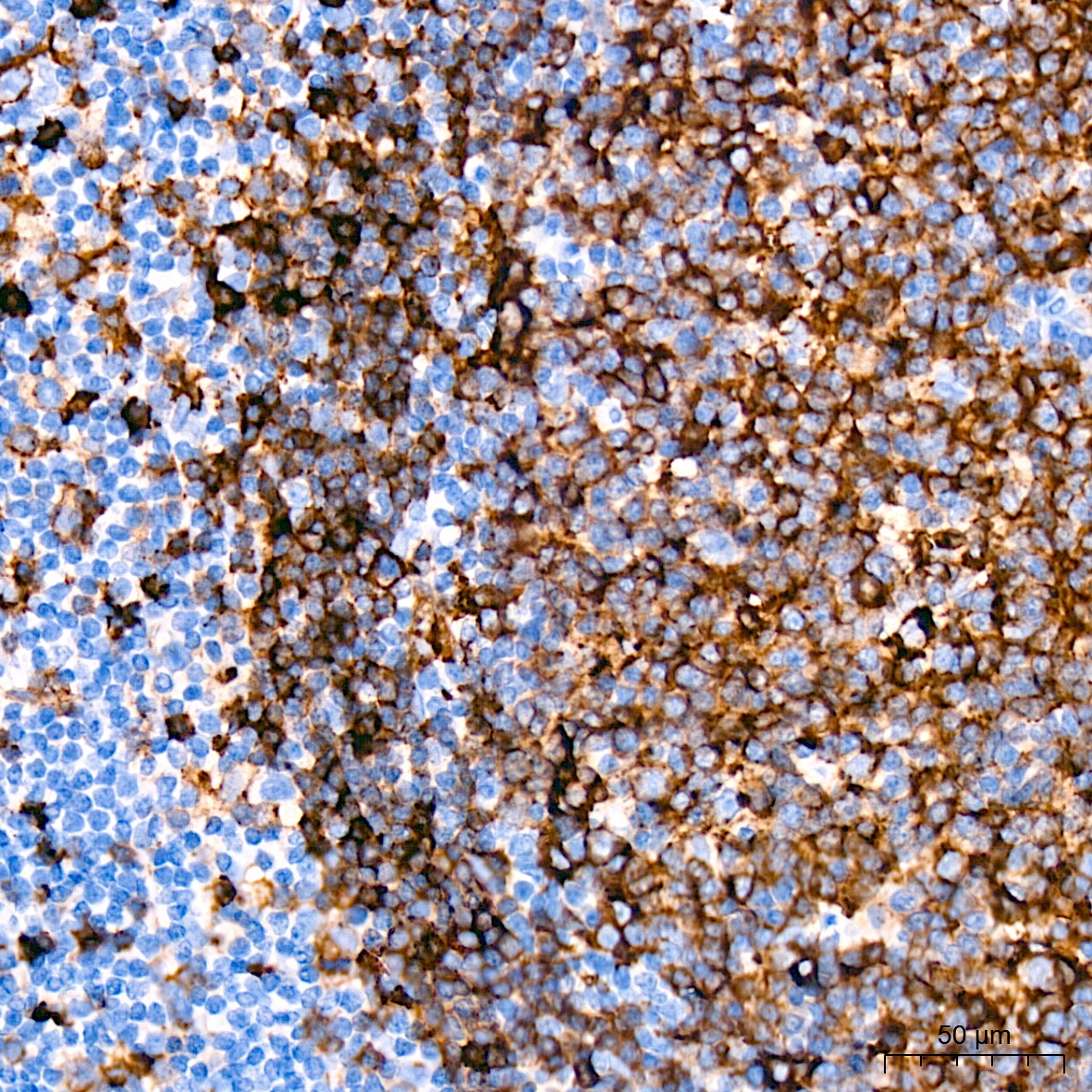
Immunohistochemistry analysis of paraffin-embedded Human tonsil tissue using HLA-DRA Rabbit mAb (CAB10863) at a dilution of 1:1600 (40x lens). High pressure antigen retrieval was performed with 0.01 M citrate buffer (pH 6.0) prior to IHC staining.
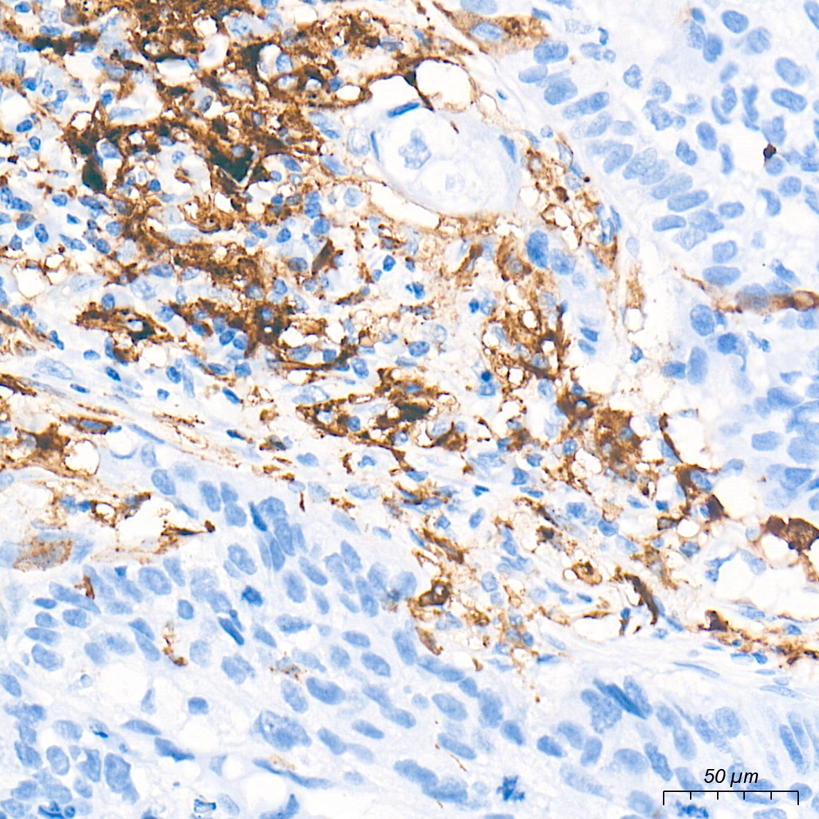
Immunohistochemistry analysis of paraffin-embedded Human lung cancer tissue using HLA-DRA Rabbit mAb (CAB10863) at a dilution of 1:2000 (40x lens). High pressure antigen retrieval performed with 0.01M Tris-EDTA Buffer (pH 9.0) prior to IHC staining.
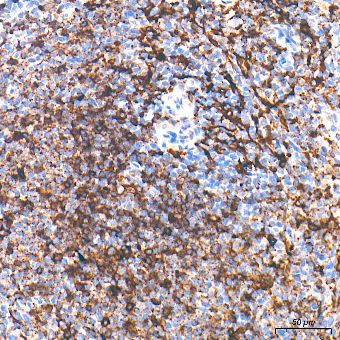
Immunohistochemistry analysis of paraffin-embedded Cynomolgus monkey spleen tissue using HLA-DRA Rabbit mAb (CAB10863) at a dilution of 1:2000 (40x lens). High pressure antigen retrieval performed with 0.01M Tris-EDTA Buffer (pH 9.0) prior to IHC staining.