The HMGA1 Monoclonal Antibody (CAB4343) is a high-quality antibody developed for reliable detection and analysis of target proteins. This antibody, generated in rabbits, exhibits high specificity and sensitivity for human samples, making it an excellent choice for various experimental applications.HMGA1 is a chromatin-associated protein that interacts with DNA and transcription factors to modulate gene expression. Dysregulation of HMGA1 has been linked to cancer development and progression, making it a promising target for therapeutic intervention.
This antibody is validated for use in WB, IHC-P, IF/ICC, ChIP-seq, ELISA applications and has demonstrated reactivity against Human, Mouse, Rat samples.
Product Name:
HMGA1 Monoclonal Antibody
SKU:
CAB4343
Size:
20μL, 100μL
Reactivity:
Human, Mouse, Rat
Clone Number:
ARC1060
Conjugate:
Unconjugated
Immunogen:
Synthetic peptide. This information is considered to be commercially sensitive.
Recommended starting concentration is 1 μg/mL. Please optimize the concentration based on your specific assay requirements.
ChIP-seq
1:50 - 1:100
Synonyms:
HMG-R, HMGIY, HMGA1A, HMGA1
Positive Sample:
HeLa, HepG2, BxPC-3, Raji, Mouse testis
Cellular Localization:
Chromosome, Nucleus.
Calculated MW:
12kDa
Observed MW:
18kDa
This gene encodes a chromatin-associated protein involved in the regulation of gene transcription, integration of retroviruses into chromosomes, and the metastatic progression of cancer cells. The encoded protein preferentially binds to the minor groove of AT-rich regions in double-stranded DNA. Multiple transcript variants encoding different isoforms have been found for this gene. Pseudogenes of this gene have been identified on multiple chromosomes.
Purification Method
Affinity purification
Gene ID
3159
RRID
AB_2863239
Buffer Information
Store at -20℃. Avoid freeze / thaw cycles. Buffer: PBS containing 50% glycerol and 0.05% BSA, preserved with proclin300 or sodium azide, pH 7.3.
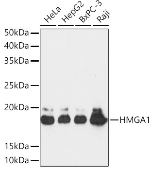
Western blot analysis of various lysates using HMGA1 Rabbit mAb (CAB4343) at 1:1000 dilution. Secondary antibody: HRP-conjugated Goat anti-Rabbit IgG (H+L) (CABS014) at 1:10000 dilution. Lysates/proteins: 25μg per lane. Blocking buffer: 3% nonfat dry milk in TBST. Detection: ECL Basic Kit (AbGn00020). Exposure time: 30s.
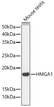
Western blot analysis of lysates from Mouse testis, using HMGA1 Rabbit mAb (CAB4343) at 1:1000 dilution. Secondary antibody: HRP-conjugated Goat anti-Rabbit IgG (H+L) (CABS014) at 1:10000 dilution. Lysates/proteins: 25μg per lane. Blocking buffer: 3% nonfat dry milk in TBST. Detection: ECL Basic Kit (AbGn00020). Exposure time: 90s.
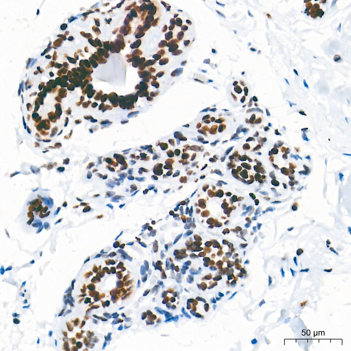
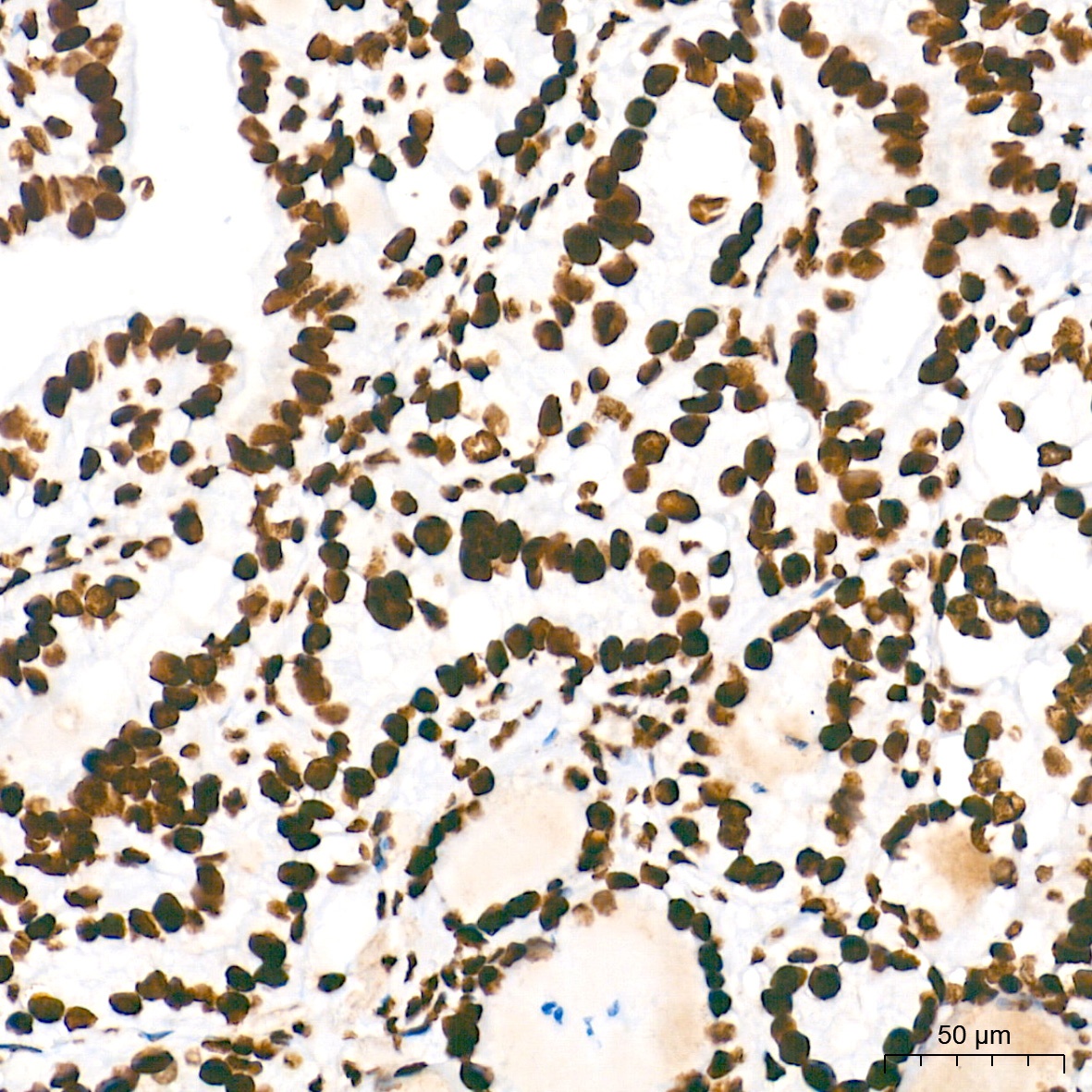
Immunohistochemistry analysis of paraffin-embedded Human breast tissue using HMGA1 Rabbit mAb (CAB4343) at a dilution of 1:5000 (40x lens). High pressure antigen retrieval performed with 0.01M Tris-EDTA Buffer (pH 9.0) prior to IHC staining.
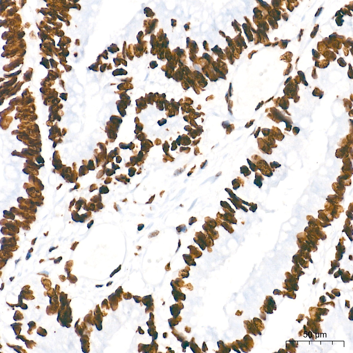
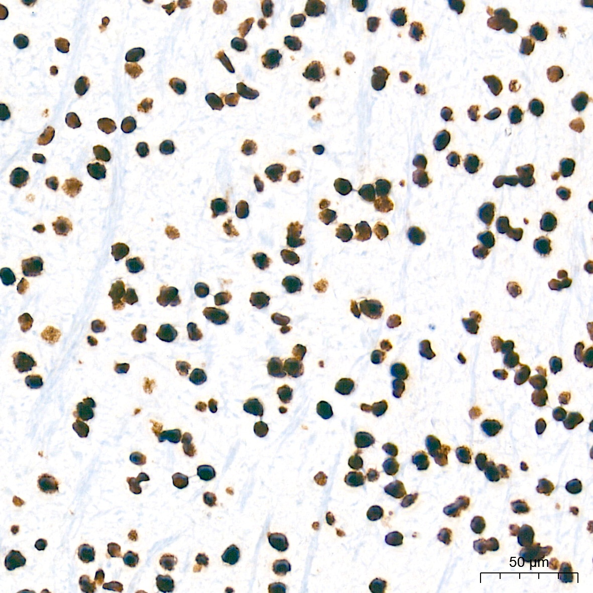
Immunohistochemistry analysis of paraffin-embedded Human colon carcinoma tissue using HMGA1 Rabbit mAb (CAB4343) at a dilution of 1:5000 (40x lens). High pressure antigen retrieval performed with 0.01M Tris-EDTA Buffer (pH 9.0) prior to IHC staining.
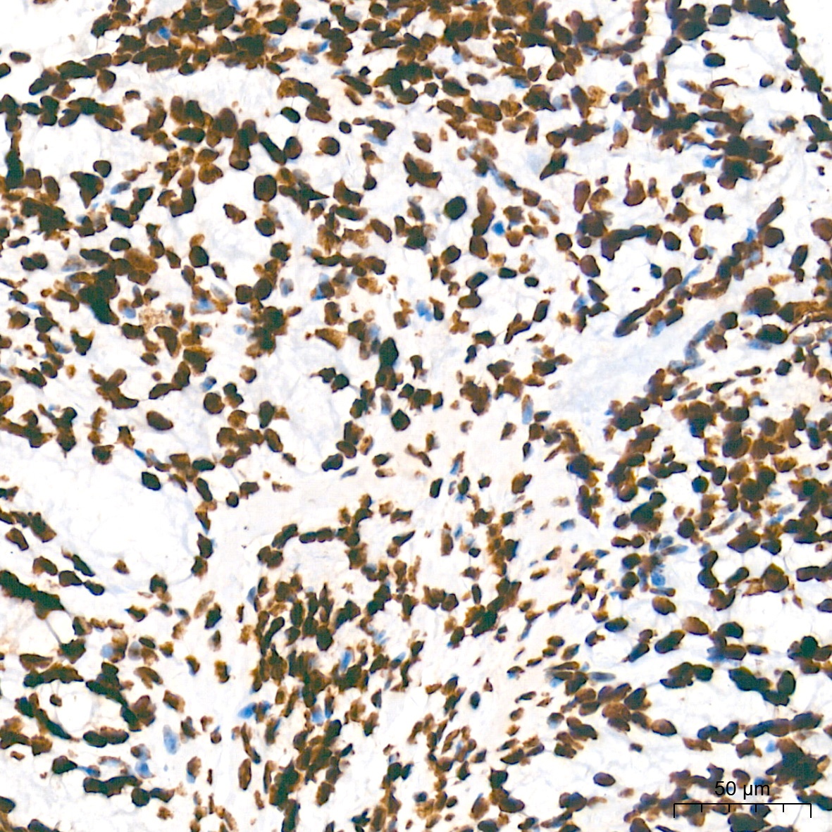
Immunohistochemistry analysis of paraffin-embedded Human thyroid cancer tissue using HMGA1 Rabbit mAb (CAB4343) at a dilution of 1:5000 (40x lens). High pressure antigen retrieval performed with 0.01M Tris-EDTA Buffer (pH 9.0) prior to IHC staining.
Immunohistochemistry analysis of paraffin-embedded Mouse brain tissue using HMGA1 Rabbit mAb (CAB4343) at a dilution of 1:5000 (40x lens). High pressure antigen retrieval performed with 0.01M Tris-EDTA Buffer (pH 9.0) prior to IHC staining.
Immunohistochemistry analysis of paraffin-embedded Mouse colon tissue using HMGA1 Rabbit mAb (CAB4343) at a dilution of 1:5000 (40x lens). High pressure antigen retrieval performed with 0.01M Tris-EDTA Buffer (pH 9.0) prior to IHC staining.
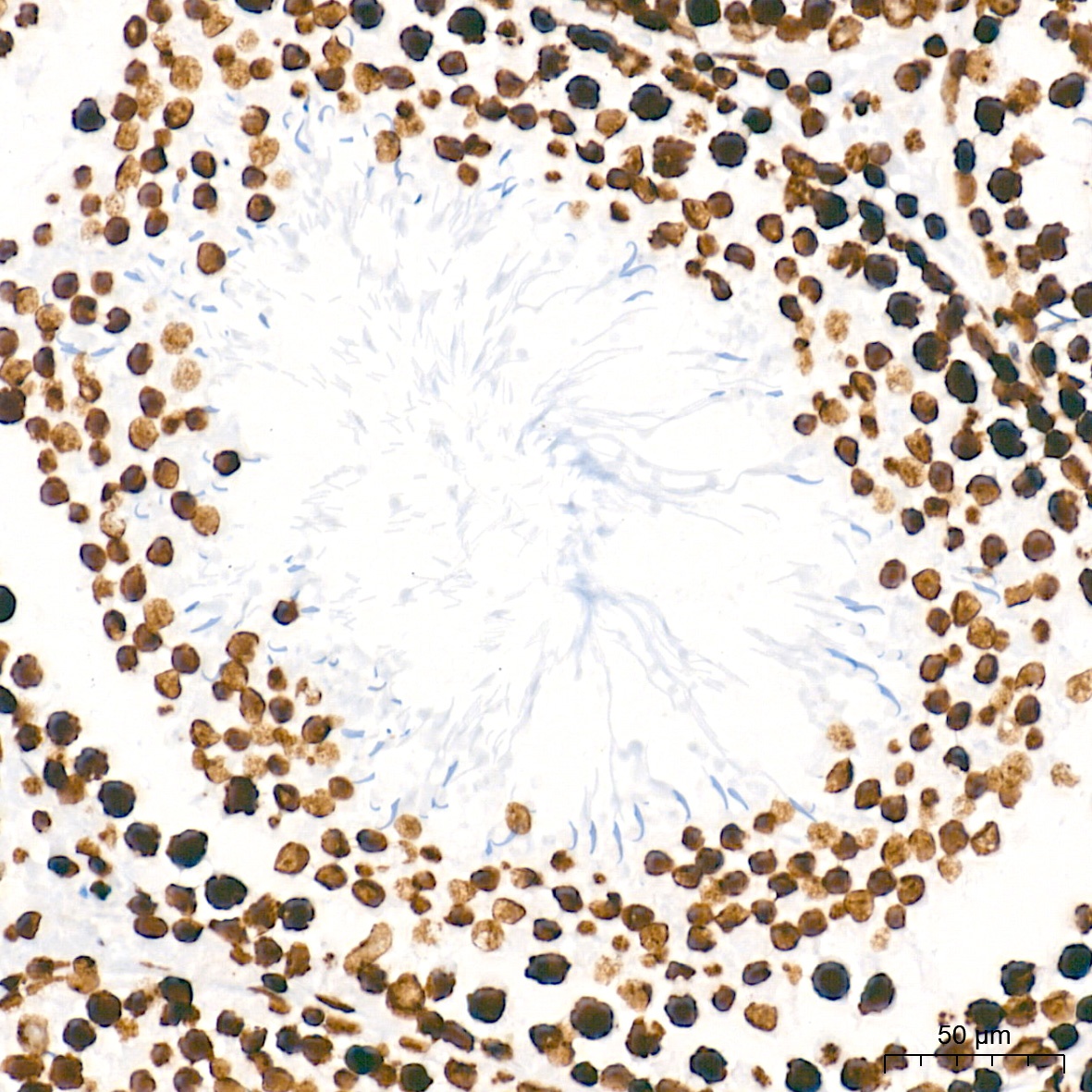
Immunohistochemistry analysis of paraffin-embedded Rat testis tissue using HMGA1 Rabbit mAb (CAB4343) at a dilution of 1:5000 (40x lens). High pressure antigen retrieval performed with 0.01M Tris-EDTA Buffer (pH 9.0) prior to IHC staining.
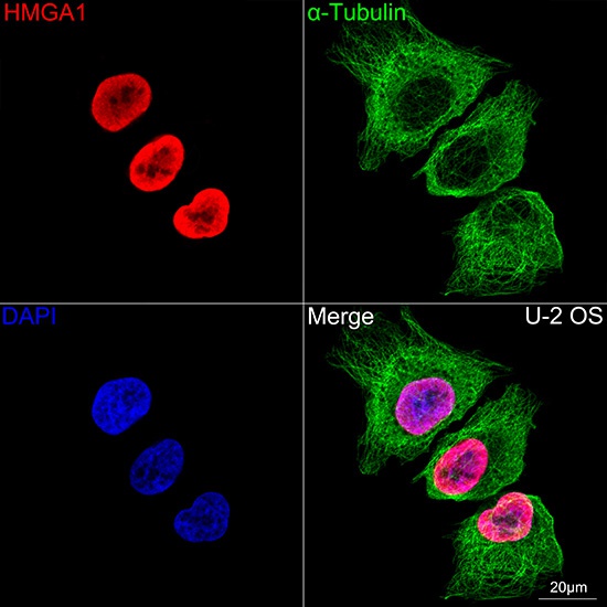
Confocal imaging of U-2 OS cells using HMGA1 Rabbit mAb (CAB4343,dilution 1:100)(Red). The cells were counterstained with α-Tubulin Mouse mAb (AC012,dilution 1:400) (Green). DAPI was used for nuclear staining (blue). Objective: 100x.