The HMGB2 Monoclonal Antibody (CAB9168) is a high-quality antibody developed for reliable detection and analysis of target proteins. This gene encodes a member of the non-histone chromosomal high mobility group protein family. The proteins of this family are chromatin-associated and ubiquitously distributed in the nucleus of higher eukaryotic cells. In vitro studies have demonstrated that this protein is able to efficiently bend DNA and form DNA circles. These studies suggest a role in facilitating cooperative interactions between cis-acting proteins by promoting DNA flexibility. This protein was also reported to be involved in the final ligation step in DNA end-joining processes of DNA double-strand breaks repair and V(D)J recombination.
This antibody is validated for use in WB, IHC-P, IF/ICC, IP, ChIP, ELISA, CUT&Tag applications and has demonstrated reactivity against Human, Mouse, Rat samples.
Product Name:
HMGB2 Monoclonal Antibody
SKU:
CAB9168
Size:
100μL, 20μL
Reactivity:
Human, Mouse, Rat
Clone Number:
ARC1460
Conjugate:
Unconjugated
Immunogen:
Synthetic peptide. This information is considered to be commercially sensitive.
Tested Applications:
WBIHC-PIF/ICCIPChIPELISACUT&Tag
Recommended Dilution:
WB
1:1000 - 1:6000
IHC-P
1:200 - 1:2000
IF/ICC
1:100 - 1:1000
IP
0.5μg-4μg antibody for 200μg-400μg extracts of whole cells
ELISA
Recommended starting concentration is 1 μg/mL. Please optimize the concentration based on your specific assay requirements.
ChIP
5μg antibody for 10μg-15μg of Chromatin
CUT&Tag
10⁵ cells /1 μg
Synonyms:
HMG2, HMGB2
Positive Sample:
HeLa, HepG2, RAW264.7, THP-1, Mouse testis, Rat testis, Rat spleen, Rat thymus,
Cellular Localization:
Chromosome, Cytoplasm, Nucleus, Secreted.
Calculated MW:
24 kDa
Observed MW:
24 kDa
This gene encodes a member of the non-histone chromosomal high mobility group protein family. The proteins of this family are chromatin-associated and ubiquitously distributed in the nucleus of higher eukaryotic cells. In vitro studies have demonstrated that this protein is able to efficiently bend DNA and form DNA circles. These studies suggest a role in facilitating cooperative interactions between cis-acting proteins by promoting DNA flexibility. This protein was also reported to be involved in the final ligation step in DNA end-joining processes of DNA double-strand breaks repair and V(D)J recombination.
Purification Method
Affinity purification
Gene ID
3148
RRID
AB_2863677
Buffer Information
Store at -20℃. Avoid freeze / thaw cycles. Buffer: PBS containing 50% glycerol and 0.05% BSA, preserved with proclin300 or sodium azide, pH 7.3.
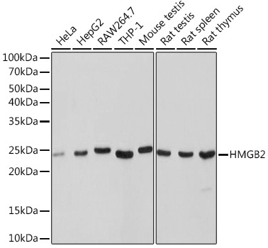
Western blot analysis of various lysates using HMGB2 Rabbit mAb (CAB9168) at 1:1000 dilution. Secondary antibody: HRP-conjugated Goat anti-Rabbit IgG (H+L) (AS014) at 1:10000 dilution. Lysates/proteins: 25μg per lane. Blocking buffer: 3% nonfat dry milk in TBST. Detection: ECL Basic Kit (AbGn00020). Exposure time: 1s.
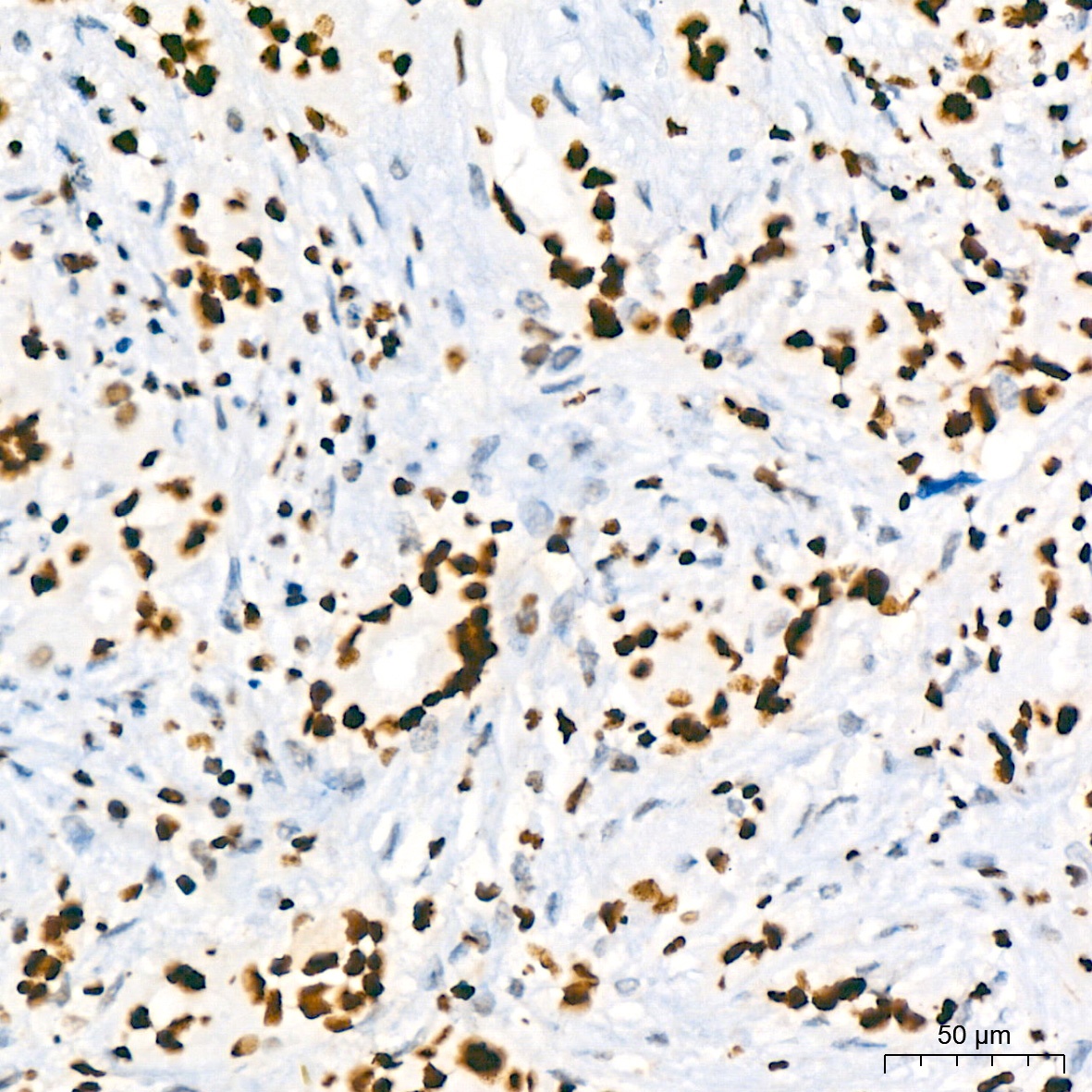
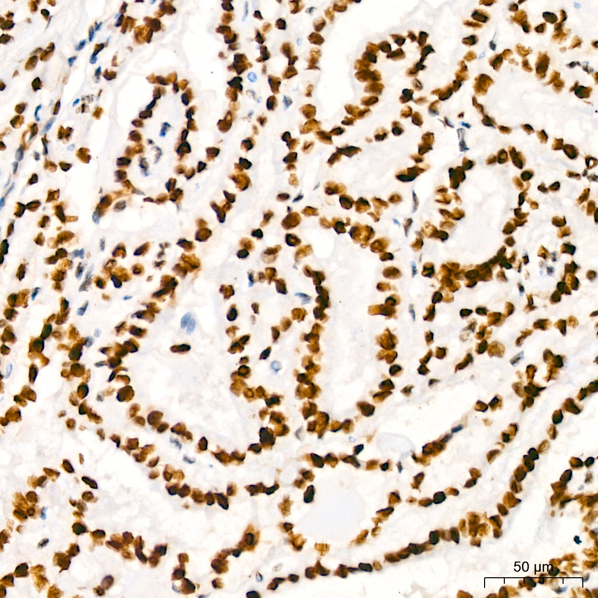
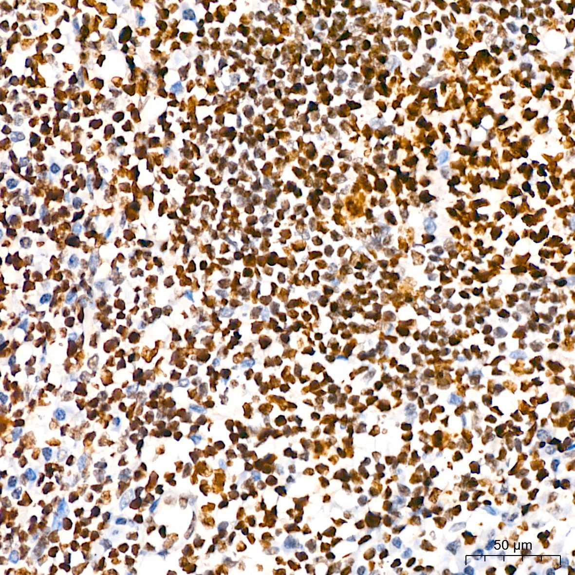
Immunohistochemistry analysis of paraffin-embedded Human breast cancer using HMGB2 Rabbit mAb (CAB9168) at dilution of 1:200 (40x lens). High pressure antigen retrieval performed with 0.01M Tris/EDTA Buffer (pH 9.0) prior to IHC staining.
Immunohistochemistry analysis of paraffin-embedded Human thyroid cancer using HMGB2 Rabbit mAb (CAB9168) at dilution of 1:200 (40x lens). High pressure antigen retrieval performed with 0.01M Tris/EDTA Buffer (pH 9.0) prior to IHC staining.
Immunohistochemistry analysis of paraffin-embedded Human tonsil using HMGB2 Rabbit mAb (CAB9168) at dilution of 1:200 (40x lens). High pressure antigen retrieval performed with 0.01M Tris/EDTA Buffer (pH 9.0) prior to IHC staining.
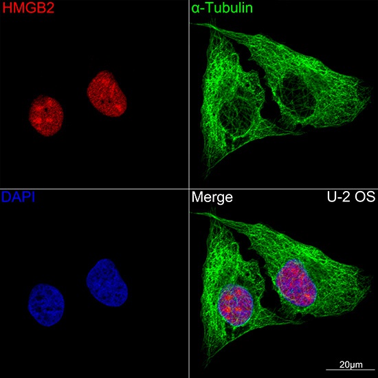
Confocal imaging of U-2 OS cells using HMGB2 Rabbit mAb (CAB9168,dilution 1:100)(Red). The cells were counterstained with α-Tubulin Mouse mAb (AC012,dilution 1:400) (Green). DAPI was used for nuclear staining (blue). Objective: 100x.
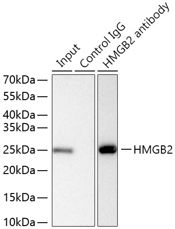
Immunoprecipitation of HMGB2 from 400 µg extracts of THP-1 cells was performed using 2 µg of HMGB2 Rabbit mAb (CAB9168). Rabbit Control IgG (AC005) was used to precipitate the Control IgG sample. IP samples were eluted with 1x Laemmli Buffer. The Input lane represents 10% of the total input. Western blot analysis of immunoprecipitates was conducted using HMGB2 Rabbit mAb (CAB9168) at a dilution of 1:1000.
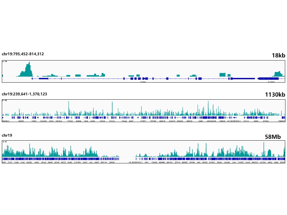
CUT&Tag was performed using the CUT&Tag Assay Kit (pAG-Tn5) for Illumina(RK20265) from 10⁵ K562 cells with 1μg HMGB2 Rabbit mAb(CAB9168), along with a Goat Anti-Rabbit IgG(H+L). The CUT&Tag results indicate the enrichment pattern of HMGB2 in representative gene loci (PTBP1), as shown in figure.