The HP1 alpha/CBX5 Monoclonal Antibody (CAB3741) is a high-quality antibody developed for reliable detection and analysis of target proteins. This antibody, raised in rabbits, exhibits high reactivity with human samples and has been validated for use in various applications, including Western blotting.HP1-alpha, also known as heterochromatin protein 1-alpha, plays a crucial role in chromatin remodeling and transcriptional regulation. Its involvement in epigenetic processes makes it a valuable target for studies in epigenetics, cancer biology, and development. The HP1-Alpha Rabbit Monoclonal Antibody enables precise detection and analysis of HP1-alpha protein levels in different experimental settings, allowing for a deeper understanding of its functions and contributions to cellular processes.
This antibody is validated for use in WB, IHC-P, IF/ICC, IP, ELISA applications and has demonstrated reactivity against Human, Mouse, Rat samples.
Product Name:
HP1 alpha/CBX5 Monoclonal Antibody
SKU:
CAB3741
Size:
20μL, 100μL
Reactivity:
Human, Mouse, Rat
Clone Number:
ARC0244
Conjugate:
Unconjugated
Immunogen:
Synthetic peptide. This information is considered to be commercially sensitive.
0.5μg-4μg antibody for 200μg-400μg extracts of whole cells
ELISA
Recommended starting concentration is 1 μg/mL. Please optimize the concentration based on your specific assay requirements.
Synonyms:
HP1, HP1A, HEL25, HP1 alpha/CBX5
Positive Sample:
HeLa, 293T, MCF7, Mouse lung, Mouse brain, Rat lung, Rat brain
Cellular Localization:
Chromosome, Nucleus, Centromere.
Calculated MW:
22kDa
Observed MW:
22kDa
This gene encodes a highly conserved nonhistone protein, which is a member of the heterochromatin protein family. The protein is enriched in the heterochromatin and associated with centromeres. The protein has a single N-terminal chromodomain which can bind to histone proteins via methylated lysine residues, and a C-terminal chromo shadow-domain (CSD) which is responsible for the homodimerization and interaction with a number of chromatin-associated nonhistone proteins. The encoded product is involved in the formation of functional kinetochore through interaction with essential kinetochore proteins. The gene has a pseudogene located on chromosome 3. Multiple alternatively spliced variants, encoding the same protein, have been identified.
Purification Method
Affinity purification
Gene ID
23468
RRID
AB_2863134
Buffer Information
Store at -20℃. Avoid freeze / thaw cycles. Buffer: PBS containing 50% glycerol and 0.05% BSA, preserved with proclin300 or sodium azide, pH 7.3.
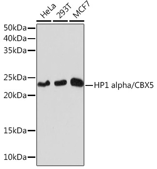
Western blot analysis of various lysates using HP1 alpha/CBX5 Rabbit mAb (CAB3741) at 1:1000 dilution incubated overnight at 4℃. Secondary antibody: HRP-conjugated Goat anti-Rabbit IgG (H+L) (CABS014) at 1:10000 dilution. Lysates/proteins: 25μg per lane. Blocking buffer: 3% nonfat dry milk in TBST. Detection: ECL Basic Kit (AbGn00020). Exposure time: 10s.
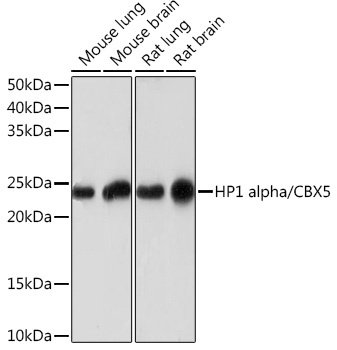
Western blot analysis of various lysates using HP1 alpha/CBX5 Rabbit mAb (CAB3741) at 1:1000 dilution incubated overnight at 4℃. Secondary antibody: HRP-conjugated Goat anti-Rabbit IgG (H+L) (CABS014) at 1:10000 dilution. Lysates/proteins: 25μg per lane. Blocking buffer: 3% nonfat dry milk in TBST. Detection: ECL Basic Kit (AbGn00020). Exposure time: 3min.
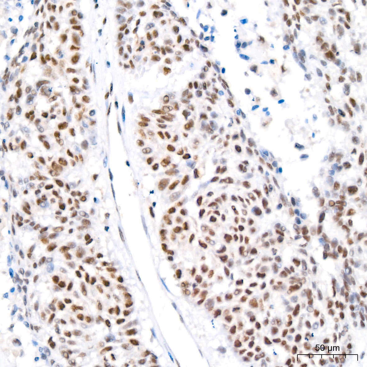
Immunohistochemistry analysis of paraffin-embedded Human lung cancer tissue using HP1 alpha/CBX5 Rabbit mAb (CAB3741) at a dilution of 1:200 (40x lens). High pressure antigen retrieval performed with 0.01M Citrate buffer (pH 6.0) prior to IHC staining.
Immunohistochemistry analysis of paraffin-embedded Human thyroid cancer tissue using HP1 alpha/CBX5 Rabbit mAb (CAB3741) at a dilution of 1:200 (40x lens). High pressure antigen retrieval performed with 0.01M Citrate buffer (pH 6.0) prior to IHC staining.
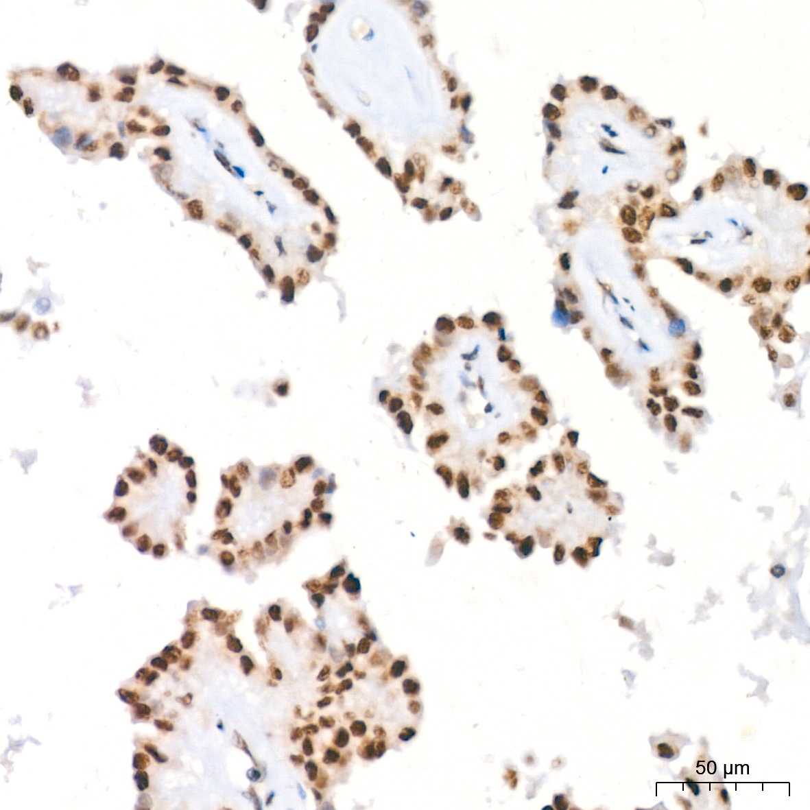
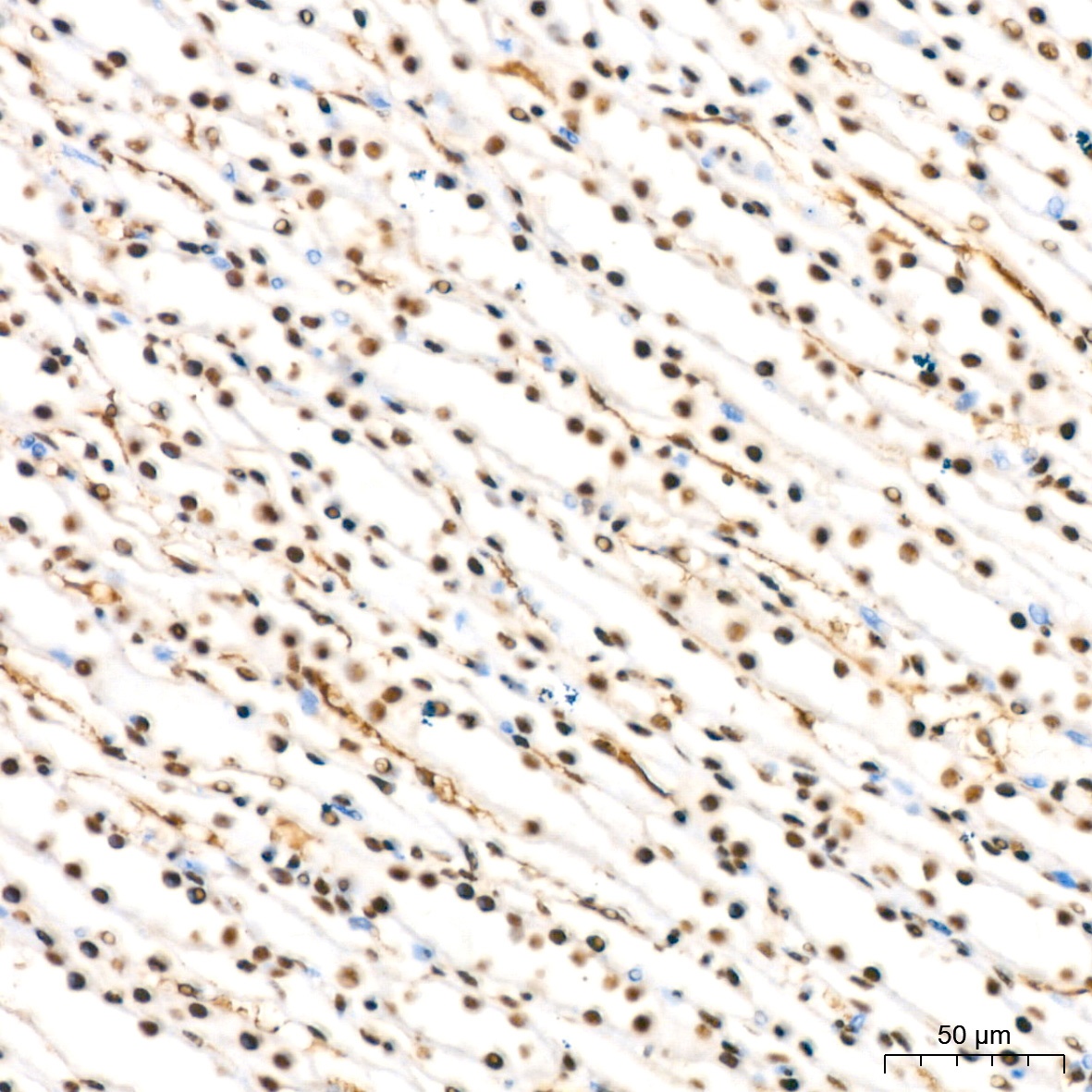
Immunohistochemistry analysis of paraffin-embedded Mouse kidney tissue using HP1 alpha/CBX5 Rabbit mAb (CAB3741) at a dilution of 1:200 (40x lens). High pressure antigen retrieval performed with 0.01M Citrate buffer (pH 6.0) prior to IHC staining.
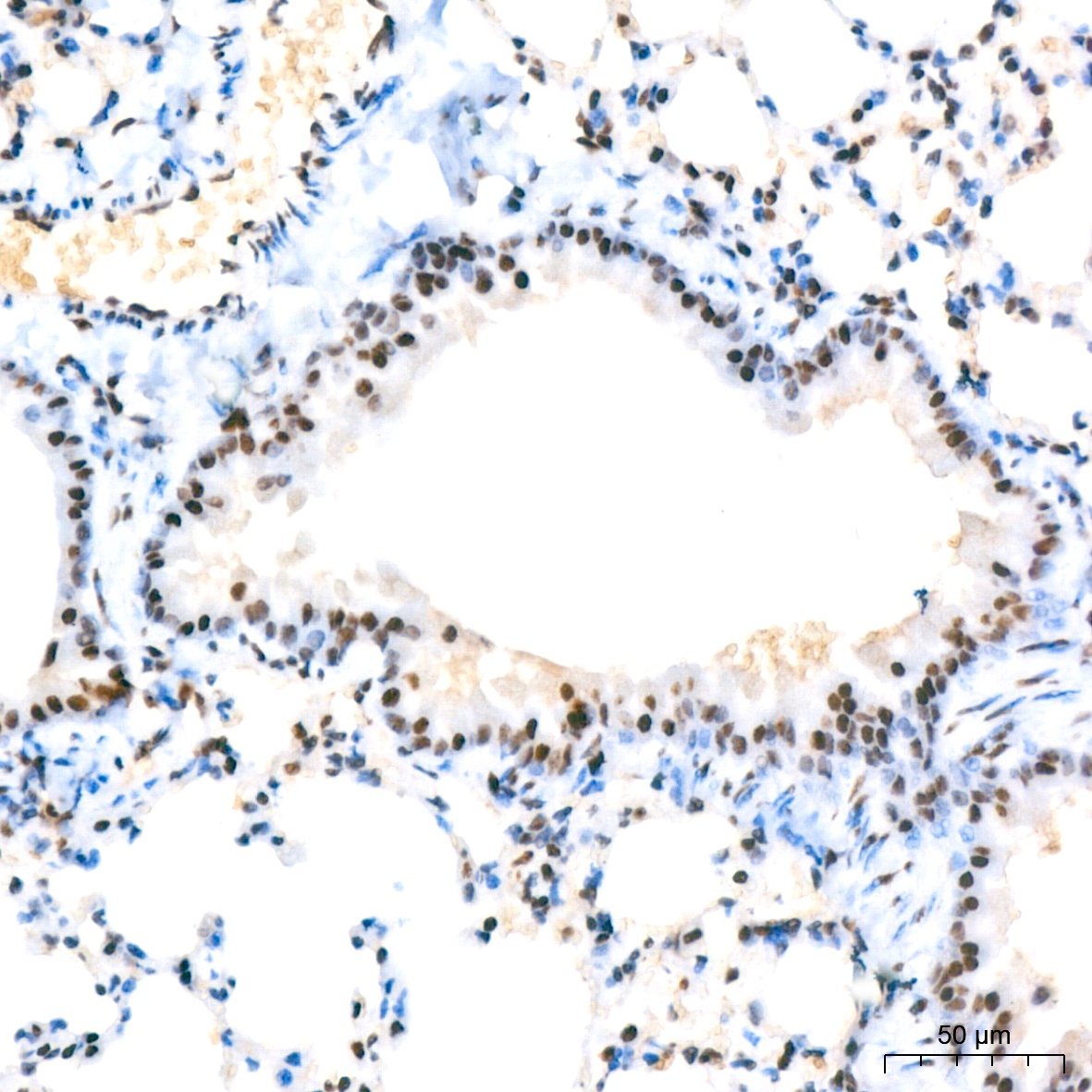
Immunohistochemistry analysis of paraffin-embedded Mouse lung tissue using HP1 alpha/CBX5 Rabbit mAb (CAB3741) at a dilution of 1:200 (40x lens). High pressure antigen retrieval performed with 0.01M Citrate buffer (pH 6.0) prior to IHC staining.
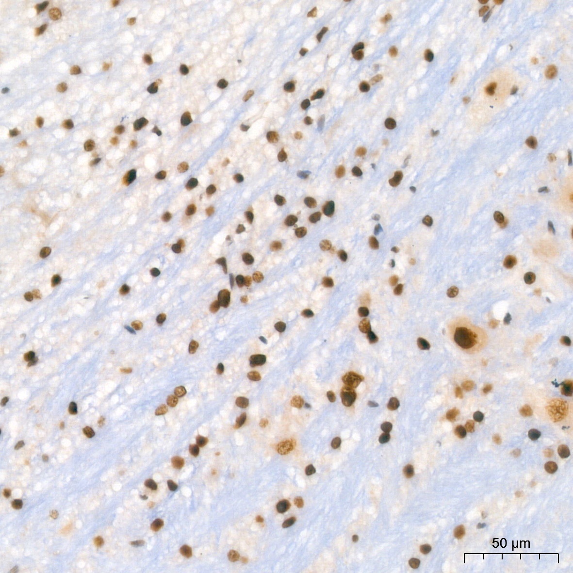
Immunohistochemistry analysis of paraffin-embedded Rat brain tissue using HP1 alpha/CBX5 Rabbit mAb (CAB3741) at a dilution of 1:200 (40x lens). High pressure antigen retrieval performed with 0.01M Citrate buffer (pH 6.0) prior to IHC staining.
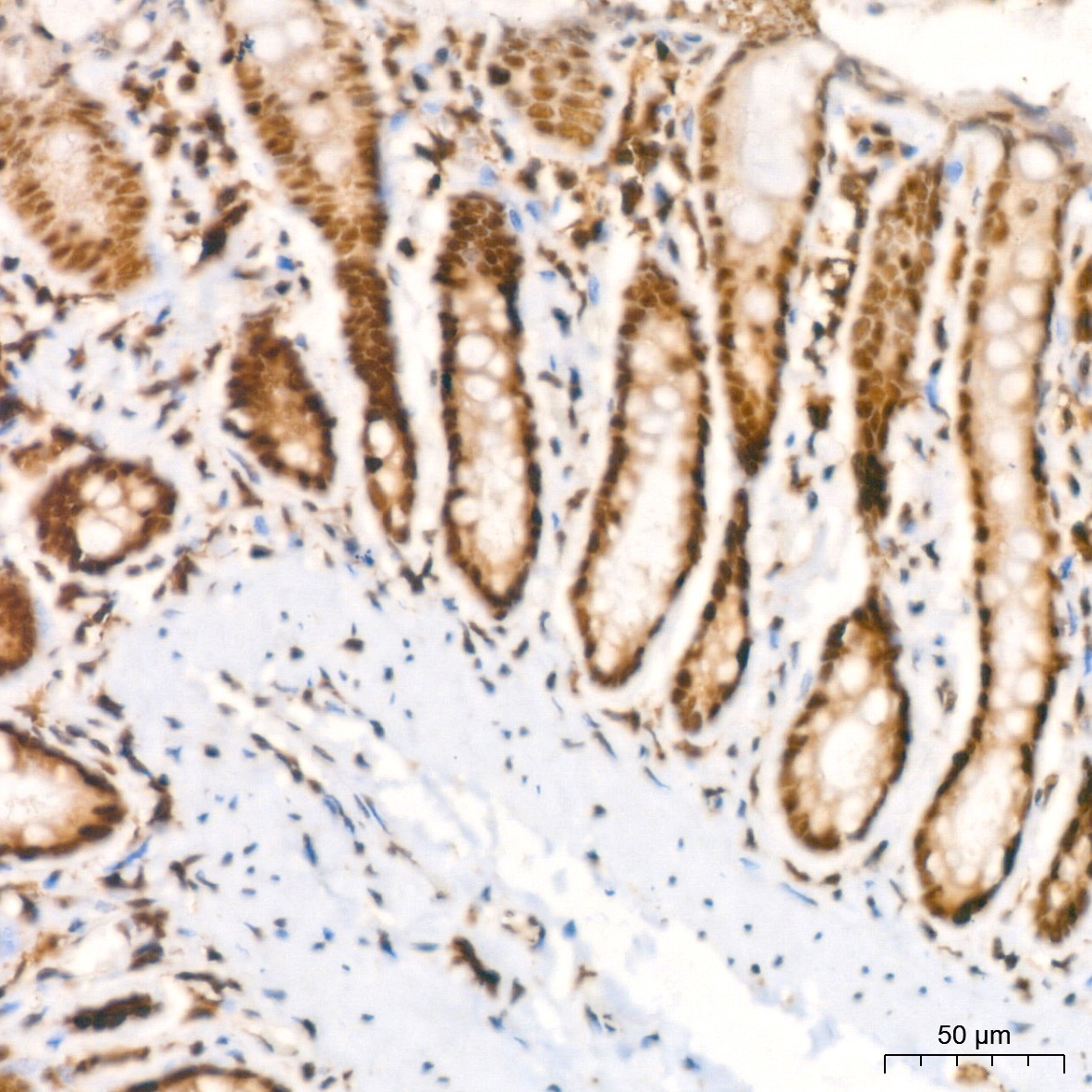
Immunohistochemistry analysis of paraffin-embedded Rat colon tissue using HP1 alpha/CBX5 Rabbit mAb (CAB3741) at a dilution of 1:200 (40x lens). High pressure antigen retrieval performed with 0.01M Citrate buffer (pH 6.0) prior to IHC staining.
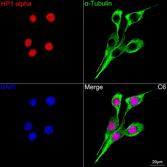
Confocal imaging of C6 cells using HP1 alpha/CBX5 Rabbit mAb (CAB3741,dilution 1:200) followed by a further incubation with Cy3 Goat Anti-Rabbit IgG (H+L) (CABS007,dilution 1:500)(Red).The cells were counterstained with α-Tubulin Mouse mAb (AC012, dilution 1:400) followed by incubation with ABflo® 488-conjugated Goat Anti-Mouse IgG (H+L) Ab (CABS076, dilution 1:500) (Green).DAPI was used for nuclear staining (Blue). Objective: 100x.


")
