")
Description
HRP-conjugated Mouse anti GFP-Tag mAb (CABE030)
The HRP-conjugated Mouse anti GFP-Tag mAb (CABE030) is a high-quality antibody developed for reliable detection and analysis of target proteins. This antibody, produced using mouse monoclonal technology, is highly sensitive and precise in detecting GFP-tagged proteins in various experimental settings.The antibody is conjugated with Horseradish Peroxidase (HRP), enabling easy and efficient detection of GFP-tagged proteins through colorimetric or chemiluminescent assays. This makes it a valuable tool for studies involving protein expression, localization, and interaction analysis.
This antibody is validated for use in WB, ELISA applications and has demonstrated reactivity against Species independent samples.
| Product Name: | HRP-conjugated Mouse anti GFP-Tag mAb |
| SKU: | CABE030 |
| Size: | 50μL, 100μL, 200μL |
| Reactivity: | Species independent |
| Clone Number: | AMC0483R-HRP |
| Conjugate: | HRP |
| Immunogen: | Synthetic peptide. This information is considered to be commercially sensitive. | ||||
| Tested Applications: | WB ELISA | ||||
| Recommended Dilution: |
| ||||
| Synonyms: | GFP, GFP tag, GFP-tag |
| Positive Sample: | insect expression of GFP |
| Observed MW: | 27kDa |
The green fluorescent protein (GFP) is a protein composed of 238 amino acid residues (26.9 kDa) that exhibits bright green fluorescence when exposed to light in the blue to ultraviolet range. Although many other marine organisms have similar green fluorescent proteins, GFP traditionally refers to the protein first isolated from the jellyfish Aequorea victoria. The GFP from A. victoria has a major excitation peak at a wavelength of 395 nm and a minor one at 475 nm. Its emission peak is at 509 nm, which is in the lower green portion of the visible spectrum. The GFP from the sea pansy (Renilla reniformis) has a single major excitation peak at 498 nm. GFP makes for an excellent tool in many forms of biology due to its ability to form internal chromophore without requiring any accessory cofactors, gene products, or enzymes / substrates other than molecular oxygen.In cell and molecular biology, the GFP gene is frequently used as a reporter of expression. It has been used in modified forms to make biosensors, and many animals have been created that express GFP, which demonstrates a proof of concept that a gene can be expressed throughout a given organism, in selected organs, or in cells of interest. GFP can be introduced into animals or other species through transgenic techniques, and maintained in their genome and that of their offspring. To date, GFP has been expressed in many species, including bacteria, yeasts, fungi, fish and mammals, including in human cells.
| Purification Method | Affinity purification |
| Buffer Information | Store at -20℃. Avoid freeze / thaw cycles. Buffer: PBS containing 50% glycerol, preserved with proclin300 or sodium azide, pH 7.3. |
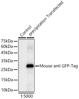 | Western blot analysis of insect expressed GFP protein using HRP-conjugated Mouse anti GFP-Tag mAb (CABE030) at 1:5000 dilution.Lysates/proteins: 25μg per lane. Blocking buffer: 3% nonfat dry milk in TBST. Detection: ECL Basic Kit (AbGn00020). Exposure time: 1s. |
")
")
")
")
")
")