The HSD17B1 Antibody (CAB10839) is a high-quality antibody developed for reliable detection and analysis of target proteins. This antibody, produced in rabbits, exhibits high reactivity with human samples and has been validated for use in Western blot applications. By specifically binding to the HSD17B1 protein, this antibody allows for the detection and analysis of HSD17B1 expression in a variety of cell types, making it a valuable asset for studies in endocrinology and hormone-related diseases.
This antibody is validated for use in WB, IHC-P, IF/ICC, ELISA applications and has demonstrated reactivity against Human, Mouse, Rat samples.
Product Name:
HSD17B1 Antibody
SKU:
CAB10839
Size:
20μL, 100μL
Reactivity:
Human, Mouse, Rat
Conjugate:
Unconjugated
Immunogen:
Recombinant protein (or fragment).This information is considered to be commercially sensitive.
This gene encodes a member of the 17beta-hydroxysteroid dehydrogenase family of short-chain dehydrogenases/reductases. It has a dual function in estrogen activation and androgen inactivation and plays a major role in establishing the estrogen E2 concentration gradient between serum and peripheral tissues. The encoded protein catalyzes the last step in estrogen activation, using NADPH to convert estrogens E1 and E2 and androgens like 4-androstenedione, to testosterone. It has an N-terminal short-chain dehydrogenase domain with a cofactor binding site, and a narrow, hydrophobic C-terminal domain with a steroid substrate binding site. This gene is expressed primarily in the placenta and ovarian granulosa cells, and to a lesser extent, in the endometrium, adipose tissue, and prostate. Polymorphisms in this gene have been linked to breast and prostate cancer. A pseudogene of this gene has been identified. Alternative splicing results in multiple transcript variants.
Purification Method
Affinity purification
Gene ID
3292
RRID
AB_2758262
Buffer Information
Store at -20℃. Avoid freeze / thaw cycles. Buffer: PBS containing 50% glycerol, preserved with proclin300 or sodium azide, pH 7.3.
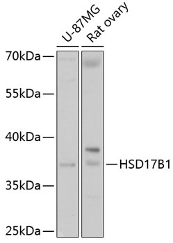
Western blot analysis of various lysates using HSD17B1 Rabbit pAb (CAB10839) at 1:1000 dilution. Secondary antibody: HRP-conjugated Goat anti-Rabbit IgG (H+L) (CABS014) at 1:10000 dilution. Lysates/proteins: 25μg per lane. Blocking buffer: 3% nonfat dry milk in TBST. Detection: ECL Basic Kit (AbGn00020). Exposure time: 5s.
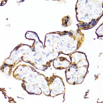
Immunohistochemistry analysis of paraffin-embedded Human placenta using HSD17B1 Rabbit pAb (CAB10839) at dilution of 1:100 (40x lens). Microwave antigen retrieval performed with 0.01M PBS Buffer (pH 7.2) prior to IHC staining.
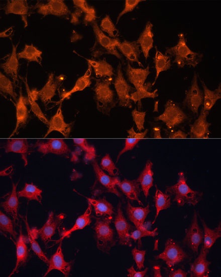
Immunofluorescence analysis of C6 cells using HSD17B1 Rabbit pAb (CAB10839) at dilution of 1:100 (40x lens). Secondary antibody: Cy3-conjugated Goat anti-Rabbit IgG (H+L) (CABS007) at 1:500 dilution. Blue: DAPI for nuclear staining.
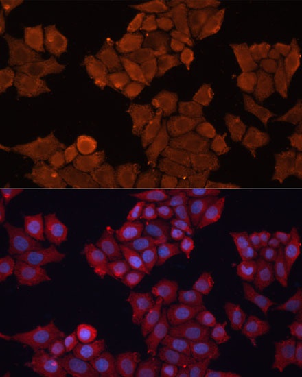
Immunofluorescence analysis of HeLa cells using HSD17B1 Rabbit pAb (CAB10839) at dilution of 1:100 (40x lens). Secondary antibody: Cy3-conjugated Goat anti-Rabbit IgG (H+L) (CABS007) at 1:500 dilution. Blue: DAPI for nuclear staining.