The HSF1 Polyclonal Antibody (CAB21994) is a high-quality antibody developed for reliable detection and analysis of target proteins. This antibody, produced in rabbits, shows high specificity for human samples and is optimized for Western blot applications. By binding specifically to HSF1, researchers can accurately detect and analyze the protein in a variety of cell types, making it an essential component for studies in cell biology, stress response, and disease mechanisms.HSF1 is a transcription factor that plays a crucial role in protecting cells from stress-induced damage, such as heat shock, oxidative stress, and protein misfolding.
This antibody is validated for use in WB, IHC-P, ELISA applications and has demonstrated reactivity against Human, Mouse samples.
Product Name:
HSF1 Polyclonal Antibody
SKU:
CAB21994
Size:
20μL, 100μL
Reactivity:
Human, Mouse
Conjugate:
Unconjugated
Immunogen:
Recombinant protein (or fragment).This information is considered to be commercially sensitive.
Recommended starting concentration is 1 μg/mL. Please optimize the concentration based on your specific assay requirements.
Synonyms:
HSF1
Positive Sample:
Mouse ovary
Cellular Localization:
Centrosome, Cytoplasm, Cytosol, Mitotic Spindle Pole, Nuclear Stress Granule, Nucleoplasm, Nucleus, Perinuclear Region Of Cytoplasm, Pml Body.
Calculated MW:
52kDa/57kDa
Observed MW:
73kDa
The product of this gene is a heat-shock transcription factor. Transcription of heat-shock genes is rapidly induced after temperature stress. Hsp90, by itself and/or associated with multichaperone complexes, is a major repressor of this gene.
Purification Method
Affinity purification
Gene ID
3297
Buffer Information
Store at -20℃. Avoid freeze / thaw cycles. Buffer: PBS containing 50% glycerol, preserved with proclin300 or sodium azide, pH 7.3.
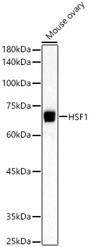
Western blot analysis of lysates from Mouse ovary, using HSF1 Rabbit pAb (CAB21994) at 1:500 dilution. Secondary antibody: HRP-conjugated Goat anti-Rabbit IgG (H+L) (CABS014) at 1:10000 dilution. Lysates/proteins: 25μg per lane. Blocking buffer: 3% nonfat dry milk in TBST. Detection: ECL Basic Kit (AbGn00020). Exposure time: 90s.
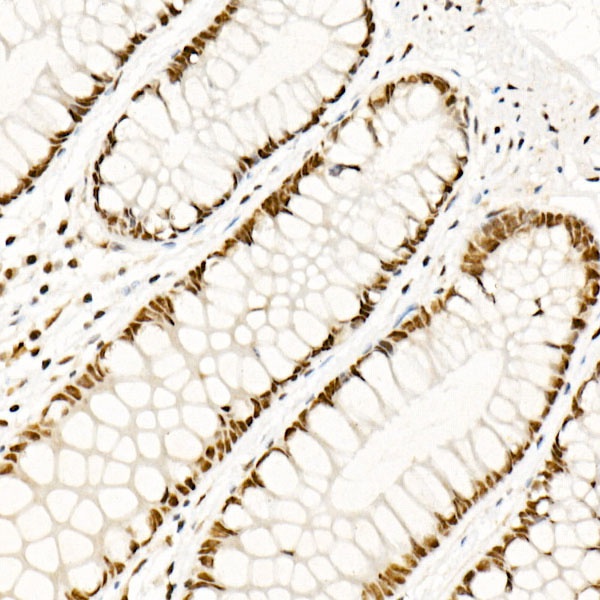
Immunohistochemistry analysis of paraffin-embedded Human colon using HSF1 Rabbit pAb (CAB21994) at dilution of 1:50 (40x lens). High pressure antigen retrieval performed with 0.01M Citrate buffer (pH 6.0) prior to IHC staining.
 at dilution of 1:50 (40x lens). Perform high pressure antigen retrieval with 10 mM citrate buffer pH 6. 0 before commencing with IHC staining protocol.")
 at dilution of 1:50 (40x lens). Perform high pressure antigen retrieval with 10 mM citrate buffer pH 6. 0 before commencing with IHC staining protocol.")


