The HSPA2 Monoclonal Antibody (CAB6652) is a high-quality antibody developed for reliable detection and analysis of target proteins. This antibody specifically targets HSPA2, a heat shock protein that plays a key role in chaperoning and protecting cellular proteins under stressful conditions. Raised in rabbits, this monoclonal antibody is highly reactive with human samples and has been validated for use in Western blot applications. By binding to the HSPA2 protein, researchers can accurately detect and analyze its expression levels in a variety of cell types. This makes the CAB6652 antibody ideal for studies in cell biology, molecular biology, and cancer research.
This antibody is validated for use in WB, IF/ICC, ELISA, IF-P applications and has demonstrated reactivity against Human, Mouse, Rat samples.
Product Name:
HSPA2 Monoclonal Antibody
SKU:
CAB6652
Size:
20μL, 100μL
Reactivity:
Human, Mouse, Rat
Clone Number:
ARC1415
Conjugate:
Unconjugated
Immunogen:
Recombinant protein (or fragment).This information is considered to be commercially sensitive.
Sequence:
ILNV TAAD KSTG KENK ITIT NDKG RLSK DDID RMVQ EAER YKSE DEAN RDRV AAKN ALES YTYN IKQT VEDE KLRG KISE QDKN KILD KCQE VINW LDRN QMAE KDEY EHKQ KELE RVCN PIIS KLYQ GGPG GGSG GGGS GASG GPTI EE
Tested Applications:
WBIF/ICCELISAIF-P
Recommended Dilution:
WB
1:500 - 1:2000
IF/ICC
1:50 - 1:200
IF-P
1:50 - 1:200
ELISA
Recommended starting concentration is 1 μg/mL. Please optimize the concentration based on your specific assay requirements.
Enables disordered domain specific binding activity; enzyme binding activity; and unfolded protein binding activity. Involved in negative regulation of inclusion body assembly and protein refolding. Located in cytosol.
Purification Method
Affinity purification
Gene ID
3306
RRID
AB_2863532
Buffer Information
Store at -20℃. Avoid freeze / thaw cycles. Buffer: PBS containing 50% glycerol and 0.05% BSA, preserved with proclin300 or sodium azide, pH 7.3.
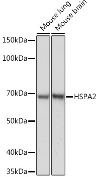
Western blot analysis of various lysates using HSPA2 Rabbit mAb (CAB6652) at 1:1000 dilution. Secondary antibody: HRP-conjugated Goat anti-Rabbit IgG (H+L) (CABS014) at 1:10000 dilution. Lysates/proteins: 25μg per lane. Blocking buffer: 3% nonfat dry milk in TBST. Detection: ECL Basic Kit (AbGn00020). Exposure time: 1s.
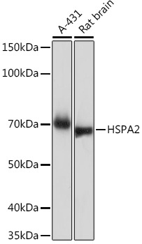
Western blot analysis of various lysates using HSPA2 Rabbit mAb (CAB6652) at 1:1000 dilution. Secondary antibody: HRP-conjugated Goat anti-Rabbit IgG (H+L) (CABS014) at 1:10000 dilution. Lysates/proteins: 25μg per lane. Blocking buffer: 3% nonfat dry milk in TBST. Detection: ECL Basic Kit (AbGn00020). Exposure time: 30s.
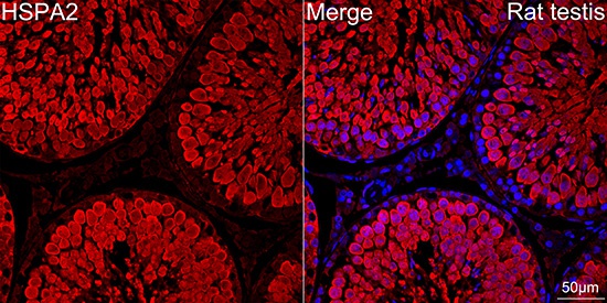
Confocal imaging of paraffin-embedded Rat testis tissue using HSPA2 Rabbit mAb (CAB6652, dilution 1:100) followed by a further incubation with Cy3 Goat Anti-Rabbit IgG (H+L) (CABS007, dilution 1:500) (Red). DAPI was used for nuclear staining (Blue). Objective: 40x. Perform high pressure antigen retrieval with 0.01 M citrate buffer (pH 6.0) prior to IF staining.