The HSP20/HSPB6 Monoclonal Antibody (CAB9091) is a high-quality antibody developed for reliable detection and analysis of target proteins. This polyclonal antibody, produced in rabbits, is highly specific and reactive with human samples, making it ideal for use in various research applications such as Western blotting.HSP20/HSPB6 is a chaperone protein that is involved in cellular stress responses and has been implicated in various diseases such as cancer, neurodegenerative disorders, and cardiovascular diseases. The Anti-HSP20/HSPB6 Antibody (CAB9091) binds specifically to the HSP20/HSPB6 protein, allowing for the detection and analysis of this important cellular component in different cell types.
This antibody is validated for use in WB, IHC-P, ELISA, IF-P applications and has demonstrated reactivity against Human, Mouse, Rat samples.
Product Name:
HSP20/HSPB6 Monoclonal Antibody
SKU:
CAB9091
Size:
20μL, 100μL
Reactivity:
Human, Mouse, Rat
Clone Number:
ARC1787
Conjugate:
Unconjugated
Immunogen:
Synthetic peptide. This information is considered to be commercially sensitive.
Recommended starting concentration is 1 μg/mL. Please optimize the concentration based on your specific assay requirements.
Synonyms:
HEL55, Hsp20, PPP1R91, HSP20/HSPB6
Positive Sample:
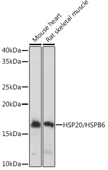
Mouse heart, Rat skeletal muscle
Cellular Localization:
Cytoplasm, Nucleus, Translocates To Nuclear Foci During Heat Shock.
Calculated MW:
17kDa
Observed MW:
17kDa
This locus encodes a heat shock protein. The encoded protein likely plays a role in smooth muscle relaxation.
Purification Method
Affinity purification
Gene ID
126393
RRID
AB_2863655
Buffer Information
Store at -20℃. Avoid freeze / thaw cycles. Buffer: PBS containing 50% glycerol and 0.05% BSA, preserved with proclin300 or sodium azide, pH 7.3.
Western blot analysis of various lysates using HSP20/HSPB6 Rabbit mAb (CAB9091) at 1:1000 dilution. Secondary antibody: HRP-conjugated Goat anti-Rabbit IgG (H+L) (CABS014) at 1:10000 dilution. Lysates/proteins: 25μg per lane. Blocking buffer: 3% nonfat dry milk in TBST. Detection: ECL Basic Kit (AbGn00020). Exposure time: 1s.
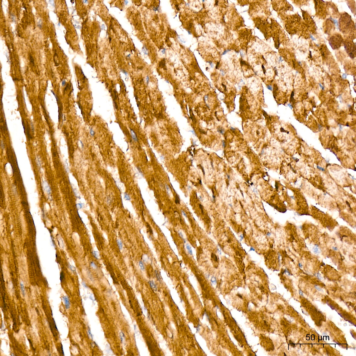
Immunohistochemistry analysis of paraffin-embedded Mouse heart tissue using HSP20/HSPB6 Rabbit mAb (CAB9091) at a dilution of 1:200 (40x lens). High pressure antigen retrieval was performed with 0.01 M Tris-EDTA buffer (pH 9.0) prior to IHC staining.
Immunohistochemistry analysis of paraffin-embedded Human liver cancer tissue using HSP20/HSPB6 Rabbit mAb (CAB9091) at a dilution of 1:200 (40x lens). High pressure antigen retrieval was performed with 0.01 M Tris-EDTA buffer (pH 9.0) prior to IHC staining.
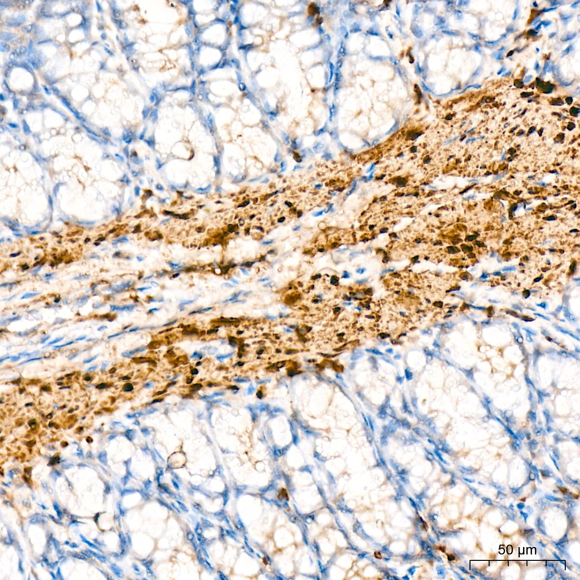
Immunohistochemistry analysis of paraffin-embedded Rat colon tissue using HSP20/HSPB6 Rabbit mAb (CAB9091) at a dilution of 1:200 (40x lens). High pressure antigen retrieval was performed with 0.01 M Tris-EDTA buffer (pH 9.0) prior to IHC staining.
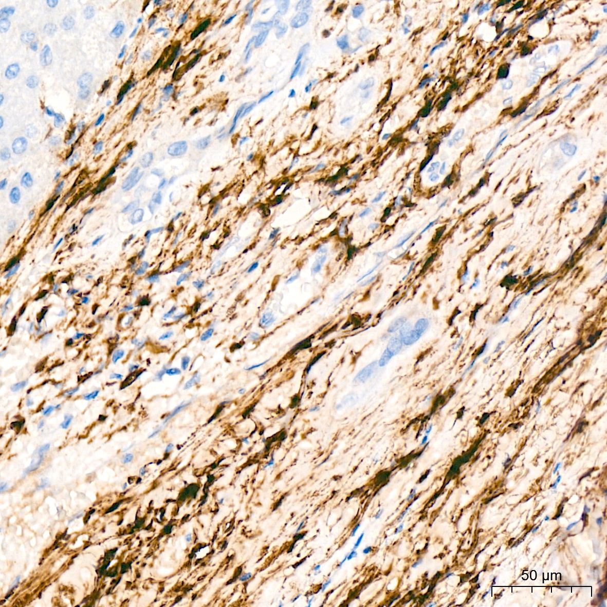
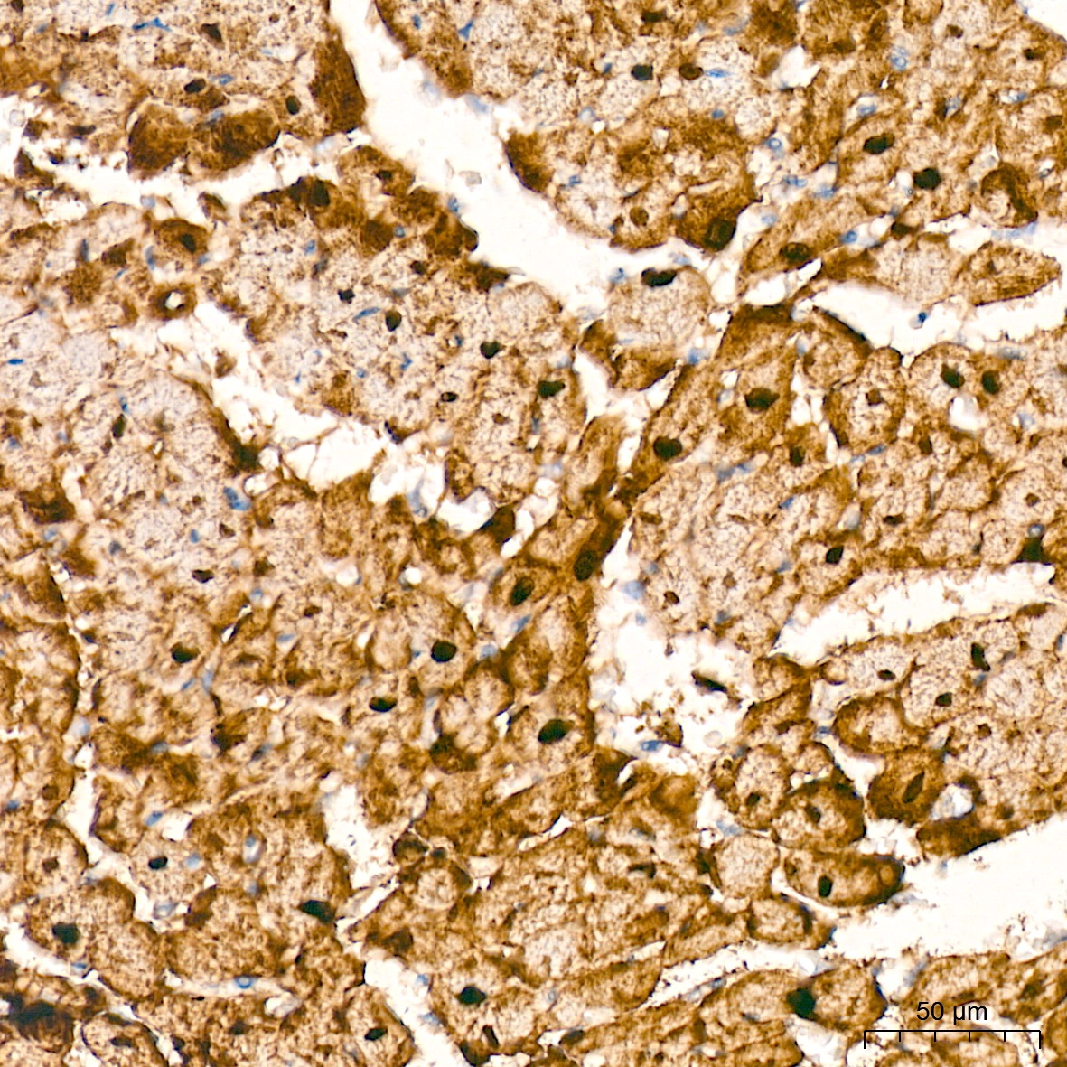
Immunohistochemistry analysis of paraffin-embedded Rat heart tissue using HSP20/HSPB6 Rabbit mAb (CAB9091) at a dilution of 1:200 (40x lens). High pressure antigen retrieval was performed with 0.01 M Tris-EDTA buffer (pH 9.0) prior to IHC staining.
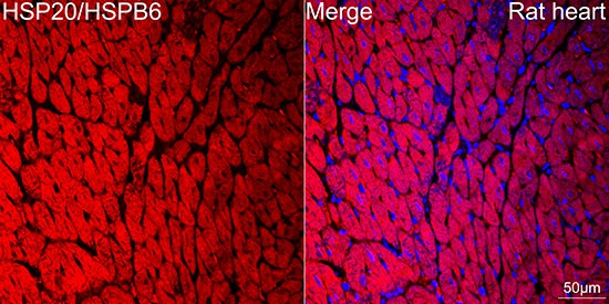
Confocal imaging of paraffin-embedded Rat heart tissue using HSP20/HSPB6 Rabbit mAb (CAB9091, dilution 1:200) followed by a further incubation with Cy3 Goat Anti-Rabbit IgG (H+L) (CABS007, dilution 1:500) (Red). DAPI was used for nuclear staining (Blue). Objective: 40x. Perform high pressure antigen retrieval with 0.01 M citrate buffer (pH 6.0) prior to IF staining.