The HSP60/HSPD1 Antibody (CAB0969) is a high-quality antibody developed for reliable detection and analysis of target proteins. This antibody, generated in rabbits, demonstrates high reactivity with human samples and has been validated for use in Western blot applications. By binding specifically to the HSPD1 protein, this antibody facilitates the detection and analysis of HSP60 in a variety of cell types.HSP60 is a chaperonin protein that plays a crucial role in the folding and assembly of other proteins within the cell. It is known to be involved in various cellular processes, including protein folding, transport, and signaling.
This antibody is validated for use in WB, IHC-P, IF/ICC, IP, ELISA applications and has demonstrated reactivity against Human, Mouse, Rat samples.
Product Name:
HSP60/HSPD1 Antibody
SKU:
CAB0969
Size:
20μL, 100μL
Reactivity:
Human, Mouse, Rat
Conjugate:
Unconjugated
Immunogen:
Recombinant protein (or fragment).This information is considered to be commercially sensitive.
This gene encodes a member of the chaperonin family. The encoded mitochondrial protein may function as a signaling molecule in the innate immune system. This protein is essential for the folding and assembly of newly imported proteins in the mitochondria. This gene is adjacent to a related family member and the region between the 2 genes functions as a bidirectional promoter. Several pseudogenes have been associated with this gene. Two transcript variants encoding the same protein have been identified for this gene. Mutations associated with this gene cause autosomal recessive spastic paraplegia 13.
Purification Method
Affinity purification
Gene ID
3329
RRID
AB_2757488
Buffer Information
Store at -20℃. Avoid freeze / thaw cycles. Buffer: PBS with 0.09% Sodium azide,50% glycerol,pH7.3.
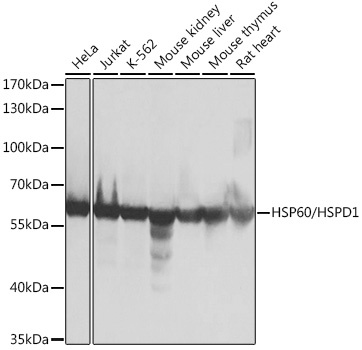
Western blot analysis of various lysates using HSP60/HSPD1 Rabbit pAb (CAB0969) at 1:1000 dilution. Secondary antibody: HRP-conjugated Goat anti-Rabbit IgG (H+L) (CABS014) at 1:10000 dilution. Lysates/proteins: 25μg per lane. Blocking buffer: 3% nonfat dry milk in TBST. Detection: ECL Basic Kit (AbGn00020). Exposure time: 10s.
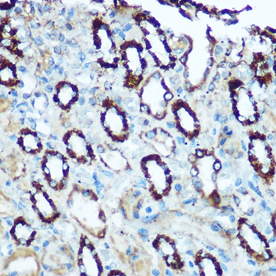
Immunohistochemistry analysis of paraffin-embedded Rat kidney using HSP60/HSPD1 Rabbit pAb (CAB0969) at dilution of 1:100 (40x lens). Microwave antigen retrieval performed with 0.01M Tris/EDTA Buffer (pH 9.0) prior to IHC staining.
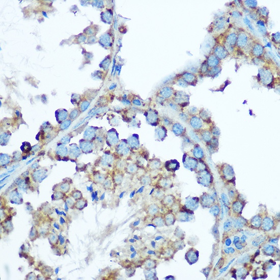
Immunohistochemistry analysis of paraffin-embedded Mouse testis using HSP60/HSPD1 Rabbit pAb (CAB0969) at dilution of 1:100 (40x lens). Microwave antigen retrieval performed with 0.01M Tris/EDTA Buffer (pH 9.0) prior to IHC staining.
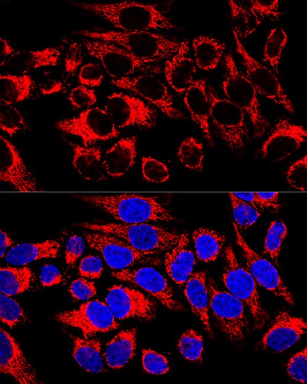
Confocal immunofluorescence analysis of HeLa cells using HSP60/HSPD1 Rabbit pAb (CAB0969) at dilution of 1:400. Blue: DAPI for nuclear staining.
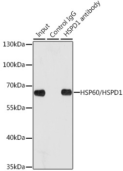
Immunoprecipitation analysis of 200 μg extracts of HeLa cells using 1 μg HSP60/HSPD1 antibody (CAB0969). Western blot was performed from the immunoprecipitate using HSP60/HSPD1 antibody (CAB0969) at a dilution of 1:1000.


 (RPES0894)")
 (RPES1972)")

