The ICOS/CD278 Antibody (CAB1811) is a high-quality antibody developed for reliable detection and analysis of target proteins. This antibody, produced in rabbits, is highly specific and reactive with human samples, making it ideal for use in Western blot applications.ICOS is a co-stimulatory molecule expressed on activated T-cells and plays a key role in regulating T-cell activation and immune response. The ICOS Polyclonal Antibody allows for the detection and analysis of ICOS protein expression in various cell types, providing valuable insights for immunological studies and cancer research.
This antibody is validated for use in WB, IF/ICC, ELISA applications and has demonstrated reactivity against Human, Mouse, Rat samples.
Product Name:
ICOS/CD278 Antibody
SKU:
CAB1811
Size:
20μL, 100μL
Reactivity:
Human, Mouse, Rat
Conjugate:
Unconjugated
Immunogen:
Recombinant protein (or fragment).This information is considered to be commercially sensitive.
Recommended starting concentration is 1 μg/mL. Please optimize the concentration based on your specific assay requirements.
Synonyms:
AILIM, CD278, CVID1, ICOS/CD278
Positive Sample:
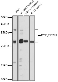
Jurkat, Mouse thymus, Mouse spleen, Rat thymus
Cellular Localization:
Cell Membrane, Secreted, Single-Pass Type I Membrane Protein.
Calculated MW:
23kDa
Observed MW:
30-40kDa
The protein encoded by this gene belongs to the CD28 and CTLA-4 cell-surface receptor family. It forms homodimers and plays an important role in cell-cell signaling, immune responses, and regulation of cell proliferation.
Purification Method
Affinity purification
Gene ID
29851
RRID
AB_2763849
Buffer Information
Store at -20℃. Avoid freeze / thaw cycles. Buffer: PBS containing 50% glycerol, preserved with proclin300 or sodium azide, pH 7.3.
Western blot analysis of various lysates using ICOS/CD278 Rabbit pAb (CAB1811) at 1:500 dilution. Secondary antibody: HRP-conjugated Goat anti-Rabbit IgG (H+L) (CABS014) at 1:10000 dilution. Lysates/proteins: 25μg per lane. Blocking buffer: 3% nonfat dry milk in TBST. Detection: ECL Basic Kit (AbGn00020). Exposure time: 180s.
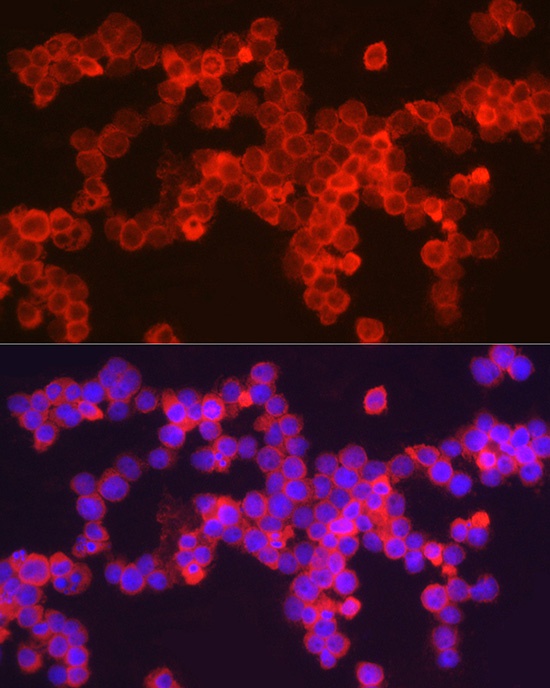
Immunofluorescence analysis of HL-60 cells using ICOS/CD278 Rabbit pAb (CAB1811) at dilution of 1:100 (40x lens). Secondary antibody: Cy3-conjugated Goat anti-Rabbit IgG (H+L) (CABS007) at 1:500 dilution. Blue: DAPI for nuclear staining.


 In Vivo Antibody - Low Endotoxin")
 In Vivo Antibody - Ultra Low Endotoxin")
")
")