The IFNAR2 Antibody (CAB1769) is a high-quality antibody developed for reliable detection and analysis of target proteins. IFNAR2 is a key player in the immune response to viral infections, as it is involved in the signaling pathway triggered by interferons. By binding specifically to IFNAR2, this antibody allows for the detection and analysis of the protein in a variety of cell types.Raised in rabbits, this antibody is highly reactive with human samples and has been validated for use in Western blot applications. Its specificity for IFNAR2 makes it an invaluable asset for studies in virology, immunology, and infectious diseases.
This antibody is validated for use in WB, IHC-P, IF/ICC, ELISA applications and has demonstrated reactivity against Human, Mouse, Rat samples.
Product Name:
IFNAR2 Antibody
SKU:
CAB1769
Size:
20μL, 100μL
Reactivity:
Human, Mouse, Rat
Conjugate:
Unconjugated
Immunogen:
Recombinant protein (or fragment).This information is considered to be commercially sensitive.
Membrane, Secreted, Single-Pass Type I Membrane Protein.
Calculated MW:
58kDa
Observed MW:
95kDa/58kDa
The protein encoded by this gene is a type I membrane protein that forms one of the two chains of a receptor for interferons alpha and beta. Binding and activation of the receptor stimulates Janus protein kinases, which in turn phosphorylate several proteins, including STAT1 and STAT2. The protein belongs to the type II cytokine receptor family. Mutations in this gene are associated with Immunodeficiency 45.
Purification Method
Affinity purification
Gene ID
3455
RRID
AB_2763812
Buffer Information
Store at -20℃. Avoid freeze / thaw cycles. Buffer: PBS containing 50% glycerol, preserved with proclin300 or sodium azide, pH 7.3.
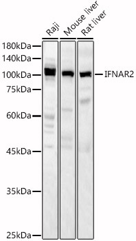
Western blot analysis of various lysates, using IFNAR2 Rabbit pAb (CAB1769) at 1:2000 dilution. Secondary antibody: HRP-conjugated Goat anti-Rabbit IgG (H+L) (CABS014) at 1:10000 dilution. Lysates/proteins: 25μg per lane. Blocking buffer: 3% nonfat dry milk in TBST. Detection: ECL Basic Kit (AbGn00020). Exposure time: 60s.
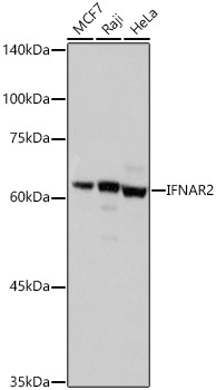
Western blot analysis of various lysates using IFNAR2 Rabbit pAb (CAB1769) at 1:500 dilution incubated overnight at 4℃. Secondary antibody: HRP-conjugated Goat anti-Rabbit IgG (H+L) (CABS014) at 1:10000 dilution. Lysates/proteins: 25 μg per lane. Blocking buffer: 3% nonfat dry milk in TBST. Detection: ECL Basic Kit (AbGn00020). Exposure time: 1s.
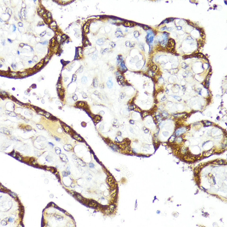
Immunohistochemistry analysis of paraffin-embedded Human placenta using IFNAR2 Rabbit pAb (CAB1769) at dilution of 1:100 (40x lens). Microwave antigen retrieval performed with 0.01M PBS Buffer (pH 7.2) prior to IHC staining.
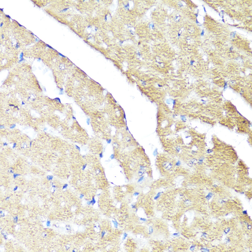
Immunohistochemistry analysis of paraffin-embedded Mouse heart using IFNAR2 Rabbit pAb (CAB1769) at dilution of 1:100 (40x lens). Microwave antigen retrieval performed with 0.01M PBS Buffer (pH 7.2) prior to IHC staining.
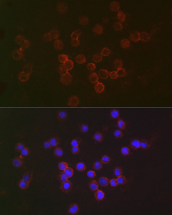
Immunofluorescence analysis of Raji cells using IFNAR2 Rabbit pAb (CAB1769) at dilution of 1:50 (40x lens). Secondary antibody: Cy3-conjugated Goat anti-Rabbit IgG (H+L) (CABS007) at 1:500 dilution. Blue: DAPI for nuclear staining.
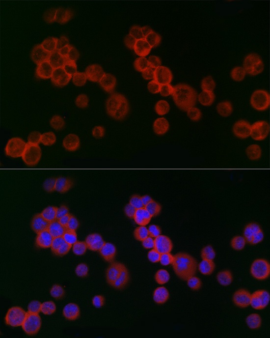
Immunofluorescence analysis of K-562 cells using IFNAR2 Rabbit pAb (CAB1769) at dilution of 1:100 (40x lens). Secondary antibody: Cy3-conjugated Goat anti-Rabbit IgG (H+L) (CABS007) at 1:500 dilution. Blue: DAPI for nuclear staining.