The IGFBP4 Antibody (CAB2008) is a high-quality antibody developed for reliable detection and analysis of target proteins. This antibody, produced in rabbits, is highly specific for human samples and is suitable for use in Western blotting applications. By binding to IGFBP4, this antibody enables the detection and analysis of IGFBP4 protein levels in various cell types, making it an essential reagent for studies in cancer biology, developmental biology, and endocrinology.IGFBP4 is known to interact with insulin-like growth factors and modulate their activity, influencing processes such as cell proliferation, differentiation, and apoptosis.
This antibody is validated for use in WB, IHC-P, IF/ICC, ELISA applications and has demonstrated reactivity against Human, Mouse, Rat samples.
Product Name:
IGFBP4 Antibody
SKU:
CAB2008
Size:
20μL, 100μL
Reactivity:
Human, Mouse, Rat
Conjugate:
Unconjugated
Immunogen:
Recombinant protein (or fragment).This information is considered to be commercially sensitive.
Recommended starting concentration is 1 μg/mL. Please optimize the concentration based on your specific assay requirements.
Synonyms:
BP-4, IBP4, IGFBP-4, HT29-IGFBP, IGFBP4
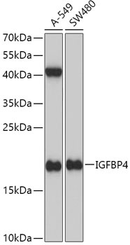
Positive Sample:
A-549, SW480
Cellular Localization:
Secreted.
Calculated MW:
28kDa
Observed MW:
17kDa
This gene is a member of the insulin-like growth factor binding protein (IGFBP) family and encodes a protein with an IGFBP domain and a thyroglobulin type-I domain. The protein binds both insulin-like growth factors (IGFs) I and II and circulates in the plasma in both glycosylated and non-glycosylated forms. Binding of this protein prolongs the half-life of the IGFs and alters their interaction with cell surface receptors.
Purification Method
Affinity purification
Gene ID
3487
RRID
AB_2764032
Buffer Information
Store at -20℃. Avoid freeze / thaw cycles. Buffer: PBS containing 50% glycerol, preserved with proclin300 or sodium azide, pH 7.3.
Western blot analysis of various lysates using IGFBP4 Rabbit pAb (CAB2008) at 1:1000 dilution. Secondary antibody: HRP-conjugated Goat anti-Rabbit IgG (H+L) (CABS014) at 1:10000 dilution. Lysates/proteins: 25μg per lane. Blocking buffer: 3% nonfat dry milk in TBST. Detection: ECL Basic Kit (AbGn00020). Exposure time: 3min.
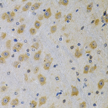
Immunohistochemistry analysis of paraffin-embedded Rat brain using IGFBP4 Rabbit pAb (CAB2008) at dilution of 1:100 (40x lens). Microwave antigen retrieval performed with 0.01M PBS Buffer (pH 7.2) prior to IHC staining.
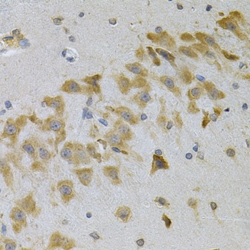
Immunohistochemistry analysis of paraffin-embedded Mouse brain using IGFBP4 Rabbit pAb (CAB2008) at dilution of 1:100 (40x lens). Microwave antigen retrieval performed with 0.01M PBS Buffer (pH 7.2) prior to IHC staining.
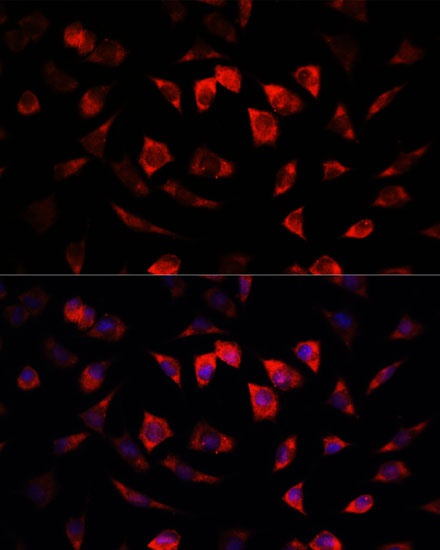
Immunofluorescence analysis of L929 cells using IGFBP4 Rabbit pAb (CAB2008) at dilution of 1:100. Secondary antibody: Cy3-conjugated Goat anti-Rabbit IgG (H+L) (CABS007) at 1:500 dilution. Blue: DAPI for nuclear staining.