The IL17A Antibody (CAB12454) is a high-quality antibody developed for reliable detection and analysis of target proteins. This gene is a member of the IL-17 receptor family which includes five members (IL-17RA-E) and the encoded protein is a proinflammatory cytokine produced by activated T cells. IL-17A-mediated downstream pathways induce the production of inflammatory molecules, chemokines, antimicrobial peptides, and remodeling proteins. The encoded protein elicits crucial impacts on host defense, cell trafficking, immune modulation, and tissue repair, with a key role in the induction of innate immune defenses. This cytokine stimulates non-hematopoietic cells and promotes chemokine production thereby attracting myeloid cells to inflammatory sites. This cytokine also regulates the activities of NF-kappaB and mitogen-activated protein kinases and can stimulate the expression of IL6 and cyclooxygenase-2 (PTGS2/COX-2), as well as enhance the production of nitric oxide (NO). IL-17A plays a pivotal role in various infectious diseases, inflammatory and autoimmune disorders, and cancer. High levels of this cytokine are associated with several chronic inflammatory diseases including rheumatoid arthritis, psoriasis and multiple sclerosis. The lung damage induced by the severe acute respiratory syndrome coronavirus 2 (SARS-CoV-2) is to a large extent, a result of the inflammatory response promoted by cytokines such as IL17A. RRID AB_2759298 Gene ID 3605 Swiss Prot Synonym IL17; CTLA8; IL-17; ILA17; CTLA-8; IL-17A; IL17A
This antibody is validated for use in WB, IHC-P, ELISA, IF-P applications and has demonstrated reactivity against Human, Mouse, Rat samples.
Product Name:
IL17A Antibody
SKU:
CAB12454
Size:
100μL, 20μL
Reactivity:
Human, Mouse, Rat
Clone Number:
-
Conjugate:
Unconjugated
Immunogen:
Recombinant protein (or fragment).This information is considered to be commercially sensitive.
Tested Applications:
WBIHC-PELISAIF-P
Recommended Dilution:
WB
1:1000 - 1:5000
IF-P
1:100 - 1:300
IHC-P
1:50 - 1:200
ELISA
Recommended starting concentration is 1 μg/mL. Please optimize the concentration based on your specific assay requirements.
Synonyms:
IL17, CTLA8, IL-17, ILA17, CTLA-8, IL-17A, IL17A
Positive Sample:
Recombinant Human IL-17A Protein
Cellular Localization:
Secreted.
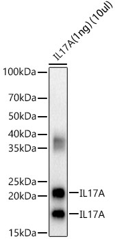
Calculated MW:
18kDa
Observed MW:
18-25kDa
This gene is a member of the IL-17 receptor family which includes five members (IL-17RA-E) and the encoded protein is a proinflammatory cytokine produced by activated T cells. IL-17A-mediated downstream pathways induce the production of inflammatory molecules, chemokines, antimicrobial peptides, and remodeling proteins. The encoded protein elicits crucial impacts on host defense, cell trafficking, immune modulation, and tissue repair, with a key role in the induction of innate immune defenses. This cytokine stimulates non-hematopoietic cells and promotes chemokine production thereby attracting myeloid cells to inflammatory sites. This cytokine also regulates the activities of NF-kappaB and mitogen-activated protein kinases and can stimulate the expression of IL6 and cyclooxygenase-2 (PTGS2/COX-2), as well as enhance the production of nitric oxide (NO). IL-17A plays a pivotal role in various infectious diseases, inflammatory and autoimmune disorders, and cancer. High levels of this cytokine are associated with several chronic inflammatory diseases including rheumatoid arthritis, psoriasis and multiple sclerosis. The lung damage induced by the severe acute respiratory syndrome coronavirus 2 (SARS-CoV-2) is to a large extent, a result of the inflammatory response promoted by cytokines such as IL17A. RRID AB_2759298 Gene ID 3605 Swiss Prot Synonym IL17; CTLA8; IL-17; ILA17; CTLA-8; IL-17A; IL17A
Purification Method:
Affinity purification
Gene ID:
3605
RRID:
AB_2759298
Buffer Information:
Store at -20℃. Avoid freeze / thaw cycles. Buffer: PBS containing 50% glycerol, preserved with proclin300 or sodium azide, pH 7.3.
Western blot analysis of various lysates, using IL17A Rabbit pAb (CAB12454) at 1:2000 dilution. Secondary antibody: HRP-conjugated Goat anti-Rabbit IgG (H+L) (AS014) at 1:10000 dilution. Lysates/proteins: 25μg per lane. Blocking buffer: 3% nonfat dry milk in TBST. Detection: ECL Basic Kit (AbGn00020). Exposure time: 60s.
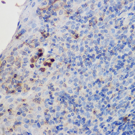
Immunohistochemistry analysis of paraffin-embedded Human tonsil using IL17A Rabbit pAb (CAB12454) at dilution of 1:200 (40x lens). High pressure antigen retrieval performed with 0.01M Citrate buffer (pH 6.0) prior to IHC staining.
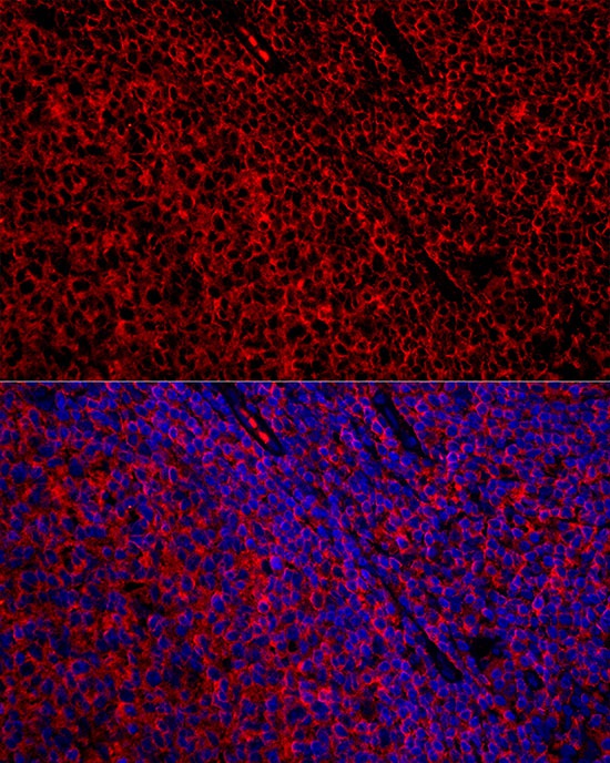
Immunofluorescence analysis of paraffin-embedded human tonsil using IL17A Rabbit pAb (CAB12454) at dilution of 1:300 (40x lens). Secondary antibody: Cy3-conjugated Goat anti-Rabbit IgG (H+L) (AS007) at 1:500 dilution. Blue: DAPI for nuclear staining.
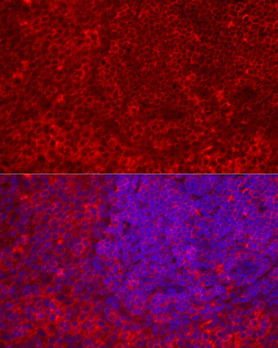
Perform high pressure antigen retrieval with 10 mM citrate buffer pH 6.0 before commencing with IF staining protocol.Immunofluorescence analysis of paraffin-embedded rat spleen using IL17A Rabbit pAb (CAB12454) at dilution of 1:300 (40x lens). Secondary antibody: Cy3-conjugated Goat anti-Rabbit IgG (H+L) (AS007) at 1:500 dilution. Blue: DAPI for nuclear staining.