The IL2 Monoclonal Antibody (CAB22200) is a high-quality antibody developed for reliable detection and analysis of target proteins. This monoclonal antibody, developed for use in various applications including ELISA, immunofluorescence, and immunohistochemistry, specifically targets IL2, allowing for precise detection and measurement in experimental settings.IL2, also known as interleukin-2, plays a vital role in the body's immune response by stimulating the proliferation of T cells and enhancing their cytotoxic activity against infected or malignant cells.
This antibody is validated for use in WB, ELISA applications and has demonstrated reactivity against Mouse samples.
Product Name:
IL2 Monoclonal Antibody
SKU:
CAB22200
Size:
20μL, 100μL
Reactivity:
Mouse
Clone Number:
ARC55614
Conjugate:
Unconjugated
Immunogen:
Recombinant protein (or fragment).This information is considered to be commercially sensitive.
Recommended starting concentration is 1 μg/mL. Please optimize the concentration based on your specific assay requirements.
Synonyms:
Il-2, IL2
Positive Sample:
C57BL/6 splenocytes treated with PMA, ionomycin and monensin; Recombinant Mouse IL-2 protein
Cellular Localization:
Extracellular Space.
Calculated MW:
19kDa
Observed MW:
22kDa(RecombinantProtein)/19kDa
Enables cytokine activity and interleukin-2 receptor binding activity. Involved in negative regulation of T-helper 17 cell differentiation. Acts upstream of or within several processes, including extrinsic apoptotic signaling pathway in absence of ligand; positive regulation of macromolecule metabolic process; and regulation of lymphocyte activation. Located in extracellular space. Is expressed in liver; omental bursa; thymus; and thymus primordium. Used to study Sjogren's syndrome and inflammatory bowel disease. Human ortholog(s) of this gene implicated in several diseases, including Takayasu's arteritis; auditory system disease (multiple); autoimmune disease (multiple); carcinoma (multiple); and neurodegenerative disease (multiple). Orthologous to human IL2 (interleukin 2).
Purification Method
Affinity purification
Gene ID
16183
Buffer Information
Store at -20℃. Avoid freeze / thaw cycles. Buffer: PBS with 0.09% Sodium azide,0.05% BSA,50% glycerol,pH7.3.
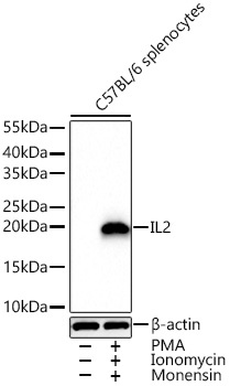
Western blot analysis of lysates from C57BL/6 splenocytes using IL2 Rabbit mAb (CAB22200) at 1:1000 dilution incubated overnight at 4℃. C57BL/6 splenocytes treated with PMA (50 ng/mL) , ionomycin (1ug/mL) at 37℃ for 1 hour and monensin (2 uM) at 37℃ for 4 hours. Secondary antibody: HRP-conjugated Goat anti-Rabbit IgG (H+L) (CABS014) at 1:10000 dilution. Lysates/proteins: 30 μg per lane. Blocking buffer: 3% nonfat dry milk in TBST. Detection: ECL Basic Kit (AbGn00020). Exposure time: 20 s.
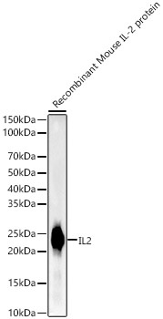
Western blot analysis of Recombinant Mouse IL-2 Protein (RP01384), using IL2 Rabbit mAb (CAB22200) at1:2000 dilution. Secondary antibody: HRP-conjugated Goat anti-Rabbit IgG (H+L) (CABS014) at 1:10000 dilution. Lysates/proteins: 10ng per lane. Blocking buffer: 3% nonfat dry milk in TBST. Detection: ECL Basic Kit (AbGn00020). Exposure time: 0.2s.
 at1:2000 dilution. Secondary antibody: HRP Goat Anti-Rabbit IgG (H+L) at 1:10000 dilution. Lysates/proteins: 25μg per lane. Blocking buffer: 3% nonfat dry milk in TBST.")
 at1:2000 dilution. Secondary antibody: HRP Goat Anti-Rabbit IgG (H+L) at 1:10000 dilution. Lysates/proteins: 25μg per lane. Blocking buffer: 3% nonfat dry milk in TBST.")


