The IL2 Antibody (CAB16317) is a high-quality antibody developed for reliable detection and analysis of target proteins. Raised in rabbits, this antibody demonstrates high reactivity with human samples and has been validated for use in Western blot applications. By specifically binding to the IL2 protein, this antibody allows for the detection and analysis of IL-2 in various cell types, making it an essential component for studies in immunology and cancer research.IL-2 is a critical factor in the proliferation and differentiation of T cells, key players in the immune response.
This antibody is validated for use in WB, IHC-P, ELISA applications and has demonstrated reactivity against Human, Mouse, Rat samples.
Product Name:
IL2 Antibody
SKU:
CAB16317
Size:
20μL, 100μL
Reactivity:
Human, Mouse, Rat
Conjugate:
Unconjugated
Immunogen:
Recombinant protein (or fragment).This information is considered to be commercially sensitive.
Recommended starting concentration is 1 μg/mL. Please optimize the concentration based on your specific assay requirements.
Synonyms:
IL-2, TCGF, lymphokine, IL2
Positive Sample:
Recombinat Human IL2 Protein, Jurkat treated with TPA , A23187 and Brefeldin A
Cellular Localization:
Secreted.
Calculated MW:
18kDa
Observed MW:
15kDa/16kDa/18kDa
This gene is a member of the interleukin 2 (IL2) cytokine subfamily which includes IL4, IL7, IL9, IL15, IL21, erythropoietin, and thrombopoietin. The protein encoded by this gene is a secreted cytokine produced by activated CD4+ and CD8+ T lymphocytes, that is important for the proliferation of T and B lymphocytes. The receptor of this cytokine (IL2R) is a heterotrimeric protein complex whose gamma chain is also shared by IL4 and IL7. The expression of this gene in mature thymocytes is monoallelic, which represents an unusual regulatory mode for controlling the precise expression of a single gene. The targeted disruption of a similar gene in mice leads to ulcerative colitis-like disease, which suggests an essential role of this gene in the immune response to antigenic stimuli.
Purification Method
Affinity purification
Gene ID
3558
RRID
AB_2769950
Buffer Information
Store at -20℃. Avoid freeze / thaw cycles. Buffer: PBS containing 50% glycerol, preserved with proclin300 or sodium azide, pH 7.3.
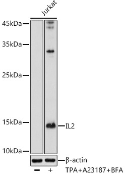
Western blot analysis of lysates from Jurkat cells, using IL2 Rabbit pAb (CAB16317) at 1:1000 dilution. Jurkat cells were treated with TPA (40 nM),A23187 (2μM) and Brefeldin A (300 ng / ml) for 0-24hours Secondary antibody: HRP-conjugated Goat anti-Rabbit IgG (H+L) (CABS014) at 1:10000 dilution. Lysates/proteins: 25μg per lane. Blocking buffer: 3% nonfat dry milk in TBST. Detection: ECL Basic Kit (AbGn00020). Exposure time: 30s.
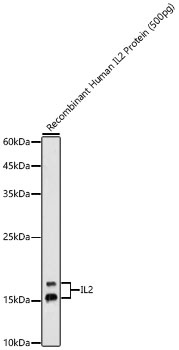
Western blot analysis of Recombinant Human IL2 Protein (RP01039), using IL2 Rabbit pAb (CAB16317) at 1:1000 dilution. Secondary antibody: HRP-conjugated Goat anti-Rabbit IgG (H+L) (CABS014) at 1:10000 dilution. Lysates/proteins: 500pg per lane. Blocking buffer: 3% nonfat dry milk in TBST. Detection: ECL Basic Kit (AbGn00020). Exposure time: 90s.
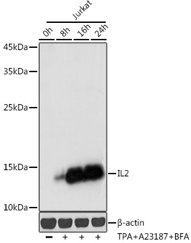
Western blot analysis of lysates from Jurkat cells, using IL2 Rabbit pAb (CAB16898) at 1:1000 dilution. Jurkat cells were treated with TPA (40 nM),A23187 (2μM) and Brefeldin A (300 ng / ml) for 0-24hours Secondary antibody: HRP-conjugated Goat anti-Rabbit IgG (H+L) (CABS014) at 1:10000 dilution. Lysates/proteins: 25μg per lane. Blocking buffer: 3% nonfat dry milk in TBST. Detection: ECL Basic Kit (AbGn00020). Exposure time: 1s.
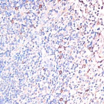
Immunohistochemistry analysis of paraffin-embedded Human tonsil using IL2 Rabbit pAb (CAB16317) at dilution of 1:100 (40x lens). Microwave antigen retrieval performed with 0.01M PBS Buffer (pH 7.2) prior to IHC staining.
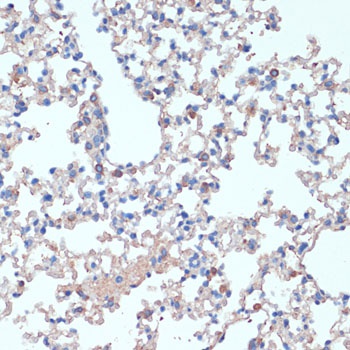
Immunohistochemistry analysis of paraffin-embedded Mouse lung using IL2 Rabbit pAb (CAB16317) at dilution of 1:100 (40x lens). Microwave antigen retrieval performed with 0.01M PBS Buffer (pH 7.2) prior to IHC staining.
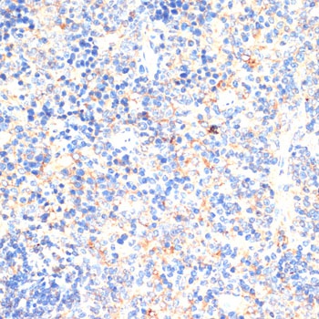
Immunohistochemistry analysis of paraffin-embedded Mouse spleen using IL2 Rabbit pAb (CAB16317) at dilution of 1:100 (40x lens). Microwave antigen retrieval performed with 0.01M PBS Buffer (pH 7.2) prior to IHC staining.