The INPP5J Antibody (CAB6631) is a high-quality antibody developed for reliable detection and analysis of target proteins. This antibody, produced in rabbits, has high specificity for human samples and has been validated for use in Western blot techniques. By targeting the INPP5J protein, this antibody allows for the detection and analysis of INPP5J in various cell types, making it suitable for studies in cell biology and signal transduction research. INPP5J is a phosphoinositide 5-phosphatase that plays a crucial role in regulating intracellular signaling cascades and membrane trafficking processes.
This antibody is validated for use in WB, IF/ICC, ELISA applications and has demonstrated reactivity against Human, Mouse, Rat samples.
Product Name:
INPP5J Antibody
SKU:
CAB6631
Size:
20μL, 100μL
Reactivity:
Human, Mouse, Rat
Conjugate:
Unconjugated
Immunogen:
Recombinant protein (or fragment).This information is considered to be commercially sensitive.
Recommended starting concentration is 1 μg/mL. Please optimize the concentration based on your specific assay requirements.
Synonyms:
PIPP, INPP5, PIB5PA, INPP5J
Positive Sample:
A-549, A375
Cellular Localization:
Cytoplasm.
Calculated MW:
107kDa
Observed MW:
107kDa
Predicted to enable phosphatidylinositol-3,4,5-trisphosphate 5-phosphatase activity and phosphatidylinositol-4,5-bisphosphate 5-phosphatase activity. Predicted to be involved in inositol phosphate dephosphorylation; negative regulation of peptidyl-serine phosphorylation; and phosphatidylinositol dephosphorylation. Predicted to act upstream of or within negative regulation of neuron projection development. Located in cytoplasm and ruffle.
Purification Method
Affinity purification
Gene ID
27124
RRID
AB_2767220
Buffer Information
Store at -20℃. Avoid freeze / thaw cycles. Buffer: PBS containing 50% glycerol, preserved with proclin300 or sodium azide, pH 7.3.
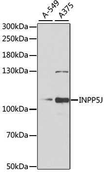
Western blot analysis of various lysates using INPP5J Rabbit pAb (CAB6631) at 1:1000 dilution. Secondary antibody: HRP-conjugated Goat anti-Rabbit IgG (H+L) (CABS014) at 1:10000 dilution. Lysates/proteins: 25μg per lane. Blocking buffer: 3% nonfat dry milk in TBST. Detection: ECL Basic Kit (AbGn00020). Exposure time: 90s.
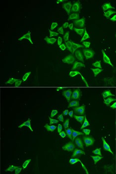
Immunofluorescence analysis of U2OS cells using INPP5J Rabbit pAb (CAB6631). Secondary antibody: Cy3-conjugated Goat anti-Rabbit IgG (H+L) (CABS007) at 1:500 dilution. Blue: DAPI for nuclear staining.